M. tuberculosis Sliding beta-Clamp Does Not Interact Directly with the NAD(+)-Dependent DNA Ligase
Kukshal, V., Khanam, T., Chopra, D., Singh, N., Sanyal, S., Ramachandran, R.(2012) PLoS One 7: e35702-e35702
- PubMed: 22545130 Search on PubMedSearch on PubMed Central
- DOI: https://doi.org/10.1371/journal.pone.0035702
- Primary Citation Related Structures:
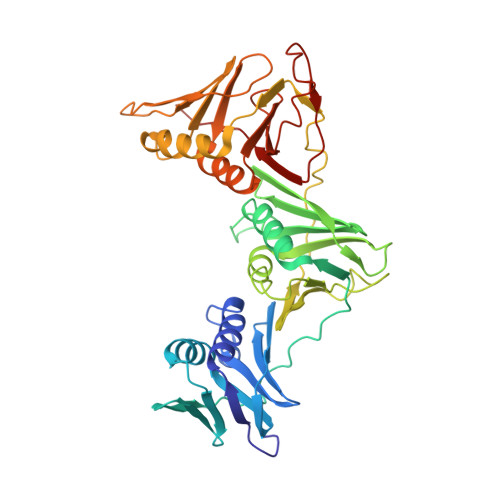
3RB9 - PubMed Abstract:
The sliding β-clamp, an important component of the DNA replication and repair machinery, is drawing increasing attention as a therapeutic target. We report the crystal structure of the M. tuberculosis β-clamp (Mtbβ-clamp) to 3.0 Å resolution. The protein crystallized in the space group C222(1) with cell-dimensions a = 72.7, b = 234.9 & c = 125.1 Å respectively. Mtbβ-clamp is a dimer, and exhibits head-to-tail association similar to other bacterial clamps. Each monomer folds into three domains with similar structures respectively and associates with its dimeric partner through 6 salt-bridges and about 21 polar interactions. Affinity experiments involving a blunt DNA duplex, primed-DNA and nicked DNA respectively show that Mtbβ-clamp binds specifically to primed DNA about 1.8 times stronger compared to the other two substrates and with an apparent K(d) of 300 nM. In bacteria like E. coli, the β-clamp is known to interact with subunits of the clamp loader, NAD(+)-dependent DNA ligase (LigA) and other partners. We tested the interactions of the Mtbβ-clamp with MtbLigA and the γ-clamp loader subunit through radioactive gel shift assays, size exclusion chromatography, yeast-two hybrid experiments and also functionally. Intriguingly while Mtbβ-clamp interacts in vitro with the γ-clamp loader, it does not interact with MtbLigA unlike in bacteria like E. coli where it does. Modeling studies involving earlier peptide complexes reveal that the peptide-binding site is largely conserved despite lower sequence identity between bacterial clamps. Overall the results suggest that other as-yet-unidentified factors may mediate interactions between the clamp, LigA and DNA in mycobacteria.
- Molecular and Structural Biology Division, Central Drug Research Institute (Council of Scientific and Industrial Research), Lucknow, Uttar Pradesh, India.
Organizational Affiliation: