3QWA | pdb_00003qwa

Crystal structure of Saccharomyces cerevisiae Zeta-crystallin-like quinone oxidoreductase Zta1


Experimental Data Snapshot

  • Method: X-RAY DIFFRACTION
  • Resolution: 2.00 Å
  • R-Value Free: 
    0.247 (Depositor), 0.247 (DCC) 
  • R-Value Work: 
    0.214 (Depositor), 0.215 (DCC) 
  • R-Value Observed: 
    0.216 (Depositor) 

Starting Model: experimental
View more details

wwPDB Validation 3D Report Full Report

Validation slider image for 3QWA

This is version 1.1 of the entry. See complete history

Literature

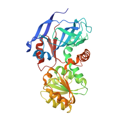
Structural insights into the cofactor-assisted substrate recognition of yeast quinone oxidoreductase Zta1

Guo, P.C.Ma, X.X.Bao, Z.Z.Ma, J.D.Chen, Y.Zhou, C.Z.

(2011) J Struct Biol 176: 112-118

  • DOI: https://doi.org/10.1016/j.jsb.2011.07.010
  • Primary Citation Related Structures: 
    3QWA, 3QWB

  • PubMed Abstract: 

    Quinone oxidoreductase (QOR EC1.6.5.5) catalyzes the reduction of quinone to hydroxyquinone using NADPH as a cofactor. Here we present the crystal structure of the ζ-crystallin-like QOR Zta1 from Saccharomycescerevisiae in apo-form at 2.00 Å and complexed with NADPH at 1.59 Å resolution. Zta1 forms a homodimer, with each subunit containing a catalytic and a cofactor-binding domain. Upon NADPH binding to the interdomain cleft, the two domains shift towards each other, producing a better fit for NADPH, and tightening substrate binding. Computational simulation combined with site-directed mutagenesis and enzymatic activity analysis defined a potential quinone-binding site that determines the stringent substrate specificity. Moreover, multiple-sequence alignment and kinetics assays implied that a single-residue change from Arg in lower organisms to Gly in vertebrates possibly resulted in elevation of enzymatic activity of ζ-crystallin-like QORs throughout evolution.


  • Organizational Affiliation
    • Hefei National Laboratory for Physical Sciences at Microscale and School of Life Sciences, University of Science and Technology of China, Hefei Anhui 230027, People's Republic of China.

Macromolecule Content 

  • Total Structure Weight: 74.13 kDa 
  • Atom Count: 5,911 
  • Modeled Residue Count: 663 
  • Deposited Residue Count: 668 
  • Unique protein chains: 1

Macromolecules

Find similar proteins by:|  3D Structure
Entity ID: 1
MoleculeChains  Sequence LengthOrganismDetailsImage
Probable quinone oxidoreductase
A, B
334Saccharomyces cerevisiae S288CMutation(s): 0 
Gene Names: YBR0421YBR046CZTA1
EC: 1.6.5.5
UniProt
Find proteins for P38230 (Saccharomyces cerevisiae (strain ATCC 204508 / S288c))
Explore P38230 
Go to UniProtKB:  P38230
Entity Groups
Sequence Clusters30% Identity50% Identity70% Identity90% Identity95% Identity100% Identity
UniProt GroupP38230
Sequence Annotations
Expand
Reference Sequence

Experimental Data & Validation

Experimental Data

  • Method: X-RAY DIFFRACTION
  • Resolution: 2.00 Å
  • R-Value Free:  0.247 (Depositor), 0.247 (DCC) 
  • R-Value Work:  0.214 (Depositor), 0.215 (DCC) 
  • R-Value Observed: 0.216 (Depositor) 
Space Group: P 21 21 21
Unit Cell:
Length ( Å )Angle ( ˚ )
a = 74.97α = 90
b = 87.313β = 90
c = 100.514γ = 90
Software Package:
Software NamePurpose
HKL-2000data collection
MOLREPphasing
REFMACrefinement
HKL-2000data reduction
HKL-2000data scaling

Structure Validation

View Full Validation Report


Entry History 

Deposition Data

Revision History  (Full details and data files)

  • Version 1.0: 2012-02-08
    Type: Initial release
  • Version 1.1: 2023-11-01
    Changes: Data collection, Database references, Refinement description