3QLZ

Candida glabrata dihydrofolate reductase complexed with NADPH and 5-[3-(2,5-dimethoxyphenyl)prop-1-yn-1-yl]-6-propylpyrimidine-2,4-diamine (UCP130B)


Experimental Data Snapshot

  • Method: X-RAY DIFFRACTION
  • Resolution: 1.94 Å
  • R-Value Free: 0.245 
  • R-Value Work: 0.206 
  • R-Value Observed: 0.208 

wwPDB Validation   3D Report Full Report


Ligand Structure Quality Assessment 


This is version 1.3 of the entry. See complete history


Literature

Crystal Structures of Candida albicans Dihydrofolate Reductase Bound to Propargyl-Linked Antifolates Reveal the Flexibility of Active Site Loop Residues Critical for Ligand Potency and Selectivity.

Paulsen, J.L.Bendel, S.D.Anderson, A.C.

(2011) Chem Biol Drug Des 78: 505-512

  • DOI: https://doi.org/10.1111/j.1747-0285.2011.01169.x
  • Primary Citation of Related Structures:  
    3QLR, 3QLS, 3QLW, 3QLX, 3QLY, 3QLZ

  • PubMed Abstract: 

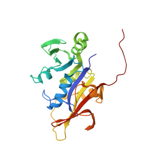
    Candida albicans and Candida glabrata cause fungal bloodstream infections that are associated with significant mortality. As part of an effort to develop potent and selective antifolates that target dihydrofolate reductase (DHFR) from Candida species, we report three ternary crystal structures of C. albicans DHFR (CaDHFR) bound to novel propargyl-linked analogs. Consistent with earlier modeling results, these structures show that hydrophobic pockets in the binding site may be exploited to increase ligand potency. The crystal structures also confirm that loop residues Thr 58- Phe 66, which flank the active site and influence ligand potency and selectivity, adopt multiple conformations. To aid the development of a dual Candida spp. inhibitor, three new crystal structures of C. glabrata DHFR (CgDHFR) bound to similar ligands as those bound in the ternary structures of CaDHFR are also reported here. Loop residues 58-66 in CgDHFR and human DHFR are 1 and 3 Å closer to the folate binding site, respectively, than loop residues in CaDHFR, suggesting that a properly size ligand could be a potent and selective dual inhibitor of CaDHFR and CgDHFR.


  • Organizational Affiliation

    Department of Pharmaceutical Sciences, University of Connecticut, Storrs, CT 06269, USA.


Macromolecules
Find similar proteins by:  (by identity cutoff)  |  3D Structure
Entity ID: 1
MoleculeChains Sequence LengthOrganismDetailsImage
Strain CBS138 chromosome J complete sequence
A, B
227Nakaseomyces glabratusMutation(s): 0 
Gene Names: CAGL0J03894gDHFR
UniProt
Find proteins for Q6FPH0 (Candida glabrata (strain ATCC 2001 / BCRC 20586 / JCM 3761 / NBRC 0622 / NRRL Y-65 / CBS 138))
Explore Q6FPH0 
Go to UniProtKB:  Q6FPH0
Entity Groups  
Sequence Clusters30% Identity50% Identity70% Identity90% Identity95% Identity100% Identity
UniProt GroupQ6FPH0
Sequence Annotations
Expand
  • Reference Sequence
Small Molecules
Binding Affinity Annotations 
IDSourceBinding Affinity
QLZ Binding MOAD:  3QLZ IC50: 15 (nM) from 1 assay(s)
Experimental Data & Validation

Experimental Data

  • Method: X-RAY DIFFRACTION
  • Resolution: 1.94 Å
  • R-Value Free: 0.245 
  • R-Value Work: 0.206 
  • R-Value Observed: 0.208 
  • Space Group: P 41
Unit Cell:
Length ( Å )Angle ( ˚ )
a = 42.607α = 90
b = 42.607β = 90
c = 230.236γ = 90
Software Package:
Software NamePurpose
SCALEPACKdata scaling
PHASERphasing
REFMACrefinement
PDB_EXTRACTdata extraction
HKL-2000data collection
DENZOdata reduction

Structure Validation

View Full Validation Report


Ligand Structure Quality Assessment 


Entry History 

Deposition Data

Revision History  (Full details and data files)

  • Version 1.0: 2011-07-20
    Type: Initial release
  • Version 1.1: 2011-09-28
    Changes: Database references
  • Version 1.2: 2013-11-20
    Changes: Non-polymer description
  • Version 1.3: 2023-09-13
    Changes: Data collection, Database references, Derived calculations, Refinement description