3QIL

Crystal structure analysis of the clathrin trimerization domain


Experimental Data Snapshot

  • Method: X-RAY DIFFRACTION
  • Resolution: 3.92 Å
  • R-Value Free: 0.386 
  • R-Value Work: 0.339 
  • R-Value Observed: 0.339 

wwPDB Validation   3D Report Full Report


This is version 1.3 of the entry. See complete history


Literature

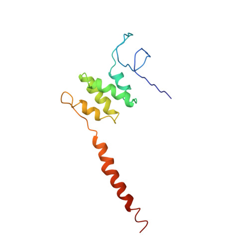
Nuclear localization of clathrin involves a labile helix outside the trimerization domain.

Ybe, J.A.Fontaine, S.N.Stone, T.Nix, J.Lin, X.Mishra, S.

(2013) FEBS Lett 587: 142-149

  • DOI: https://doi.org/10.1016/j.febslet.2012.11.005
  • Primary Citation of Related Structures:  
    3QIL

  • PubMed Abstract: 

    Clathrin is a trimeric protein involved in receptor-mediated-endocytosis, but can function as a non-trimer outside of endocytosis. We have discovered that the subcellular distribution of a clathrin cysteine mutant we previously studied is altered and a proportion is also localized to nuclear spaces. MALS shows C1573A hub is a mixture of trimer-like and detrimerized molecules. The X-ray structure of the trimerization domain reveals that without light chains, a helix harboring cysteine-1573 is reoriented. We propose clathrin has a detrimerization switch, which suggests clathrin topology can be altered naturally for new functions.


  • Organizational Affiliation

    Department of Molecular and Cellular Biochemistry, Indiana University, Bloomington, 212 S. Hawthorne Drive, Bloomington, IN 47405, USA. jybe@indiana.edu


Macromolecules
Find similar proteins by:  (by identity cutoff)  |  3D Structure
Entity ID: 1
MoleculeChains Sequence LengthOrganismDetailsImage
Clathrin heavy chain 1
A, B, C, D, E
A, B, C, D, E, F, G, H, I, J, K, L, M, N, O, P, Q, R, S, T, U, V, W, X
125Bos taurusMutation(s): 2 
Gene Names: CLTC
UniProt
Find proteins for P49951 (Bos taurus)
Explore P49951 
Go to UniProtKB:  P49951
Entity Groups  
Sequence Clusters30% Identity50% Identity70% Identity90% Identity95% Identity100% Identity
UniProt GroupP49951
Sequence Annotations
Expand
  • Reference Sequence
Experimental Data & Validation

Experimental Data

  • Method: X-RAY DIFFRACTION
  • Resolution: 3.92 Å
  • R-Value Free: 0.386 
  • R-Value Work: 0.339 
  • R-Value Observed: 0.339 
  • Space Group: H 3
Unit Cell:
Length ( Å )Angle ( ˚ )
a = 255.78α = 90
b = 255.78β = 90
c = 312.99γ = 120
Software Package:
Software NamePurpose
d*TREKdata scaling
PHASERphasing
CNSrefinement
PDB_EXTRACTdata extraction
CrystalCleardata collection
d*TREKdata reduction

Structure Validation

View Full Validation Report


Entry History 

Deposition Data

Revision History  (Full details and data files)

  • Version 1.0: 2012-02-01
    Type: Initial release
  • Version 1.1: 2013-01-16
    Changes: Database references
  • Version 1.2: 2013-06-19
    Changes: Database references
  • Version 1.3: 2024-02-21
    Changes: Data collection, Database references