3QG9 | pdb_00003qg9

crystal structure of FBF-2/gld-1 FBEa A7U mutant complex


Experimental Data Snapshot

  • Method: X-RAY DIFFRACTION
  • Resolution: 2.25 Å
  • R-Value Free: 
    0.225 (Depositor), 0.216 (DCC) 
  • R-Value Work: 
    0.170 (Depositor), 0.163 (DCC) 

wwPDB Validation 3D Report Full Report

Validation slider image for 3QG9

This is version 1.3 of the entry. See complete history

Literature

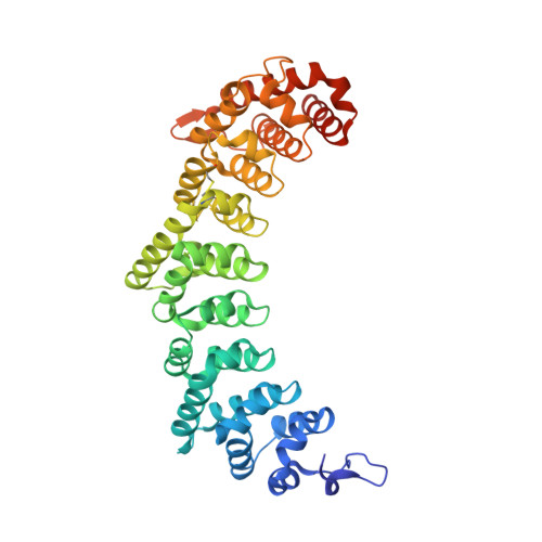
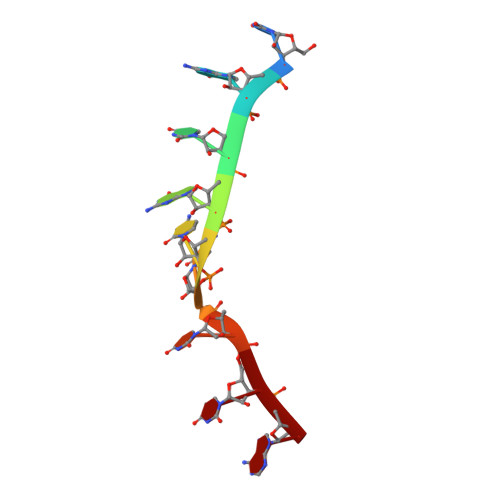
Stacking interactions in PUF-RNA complexes.

Koh, Y.Y.Wang, Y.Qiu, C.Opperman, L.Gross, L.Tanaka Hall, T.M.Wickens, M.

(2011) RNA 17: 718-727

  • DOI: https://doi.org/10.1261/rna.2540311
  • Primary Citation Related Structures: 
    3QG9, 3QGB, 3QGC

  • PubMed Abstract: 

    Stacking interactions between amino acids and bases are common in RNA-protein interactions. Many proteins that regulate mRNAs interact with single-stranded RNA elements in the 3' UTR (3'-untranslated region) of their targets. PUF proteins are exemplary. Here we focus on complexes formed between a Caenorhabditis elegans PUF protein, FBF, and its cognate RNAs. Stacking interactions are particularly prominent and involve every RNA base in the recognition element. To assess the contribution of stacking interactions to formation of the RNA-protein complex, we combine in vivo selection experiments with site-directed mutagenesis, biochemistry, and structural analysis. Our results reveal that the identities of stacking amino acids in FBF affect both the affinity and specificity of the RNA-protein interaction. Substitutions in amino acid side chains can restrict or broaden RNA specificity. We conclude that the identities of stacking residues are important in achieving the natural specificities of PUF proteins. Similarly, in PUF proteins engineered to bind new RNA sequences, the identity of stacking residues may contribute to "target" versus "off-target" interactions, and thus be an important consideration in the design of proteins with new specificities.


  • Organizational Affiliation
    • Department of Biochemistry, University of Wisconsin, Madison, Wisconsin 53706, USA.

Macromolecule Content 

  • Total Structure Weight: 49.89 kDa 
  • Atom Count: 3,664 
  • Modeled Residue Count: 409 
  • Deposited Residue Count: 422 
  • Unique protein chains: 1
  • Unique nucleic acid chains: 1

Macromolecules


Find similar proteins by:|  3D Structure
Entity ID: 1
MoleculeChains  Sequence LengthOrganismDetailsImage
Fem-3 mRNA-binding factor 2413Caenorhabditis elegansMutation(s): 0 
Gene Names: fbf-2F21H12.5
UniProt
Find proteins for Q09312 (Caenorhabditis elegans)
Explore Q09312 
Go to UniProtKB:  Q09312
Entity Groups
Sequence Clusters30% Identity50% Identity70% Identity90% Identity95% Identity100% Identity
UniProt GroupQ09312
Sequence Annotations
Expand
Reference Sequence
Find similar nucleic acids by:  Sequence
Entity ID: 2
MoleculeChains LengthOrganismImage
5'-R(*UP*GP*UP*GP*CP*CP*UP*UP*A)-3'9N/A
Sequence Annotations
Expand
Reference Sequence

Small Molecules

Ligands 1 Unique
IDChains Name / Formula / InChI Key2D Diagram3D Interactions
EDO

Query on EDO


Download:Ideal Coordinates CCD File
C [auth A]1,2-ETHANEDIOL
C2 H6 O2
LYCAIKOWRPUZTN-UHFFFAOYSA-N

Experimental Data & Validation

Experimental Data

  • Method: X-RAY DIFFRACTION
  • Resolution: 2.25 Å
  • R-Value Free:  0.225 (Depositor), 0.216 (DCC) 
  • R-Value Work:  0.170 (Depositor), 0.163 (DCC) 
Space Group: P 61
Unit Cell:
Length ( Å )Angle ( ˚ )
a = 96.75α = 90
b = 96.75β = 90
c = 101.175γ = 120
Software Package:
Software NamePurpose
HKL-2000data collection
PHASERphasing
PHENIXrefinement
HKL-2000data reduction
HKL-2000data scaling

Structure Validation

View Full Validation Report


Entry History 

Deposition Data

Revision History  (Full details and data files)

  • Version 1.0: 2011-03-23
    Type: Initial release
  • Version 1.1: 2011-07-13
    Changes: Version format compliance
  • Version 1.2: 2018-01-24
    Changes: Structure summary
  • Version 1.3: 2024-11-06
    Changes: Data collection, Database references, Derived calculations, Structure summary