Structure and Activity of Aspergillus nidulans Copper Amine Oxidase
McGrath, A.P., Mithieux, S.M., Collyer, C.A., Bakhuis, J.G., van den Berg, M., Sein, A., Heinz, A., Schmelzer, C., Weiss, A.S., Guss, J.M.(2011) Biochemistry 50: 5718-5730
- PubMed: 21604787 Search on PubMed
- DOI: https://doi.org/10.1021/bi200555c
- Primary Citation Related Structures:
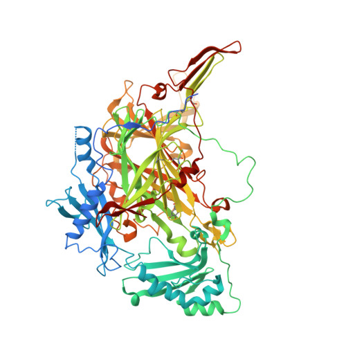
3PGB - PubMed Abstract:
Aspergillus nidulans amine oxidase (ANAO) has the unusual ability among the family of copper and trihydroxyphenylalanine quinone-containing amine oxidases of being able to oxidize the amine side chains of lysine residues in large peptides and proteins. We show here that in common with the related enzyme from the yeast Pichia pastoris, ANAO can promote the cross-linking of tropoelastin and oxidize the lysine residues in α-casein proteins and tropoelastin. The crystal structure of ANAO, the first for a fungal enzyme in this family, has been determined to a resolution of 2.4 Å. The enzyme is a dimer with the archetypal fold of a copper-containing amine oxidase. The active site is the most open of any of those of the structurally characterized enzymes in the family and provides a ready explanation for its lysine oxidase-like activity.
- School of Molecular Bioscience, University of Sydney, Sydney, NSW 2006, Australia.
Organizational Affiliation: