3PCH | pdb_00003pch

STRUCTURE OF PROTOCATECHUATE 3,4-DIOXYGENASE COMPLEXED WITH 3-CHLORO-4-HYDROXYBENZOATE


Experimental Data Snapshot

  • Method: X-RAY DIFFRACTION
  • Resolution: 2.05 Å
  • R-Value Work: 
    0.167 (Depositor) 

wwPDB Validation   3D Report Full Report


This is version 3.0 of the entry. See complete history


Literature

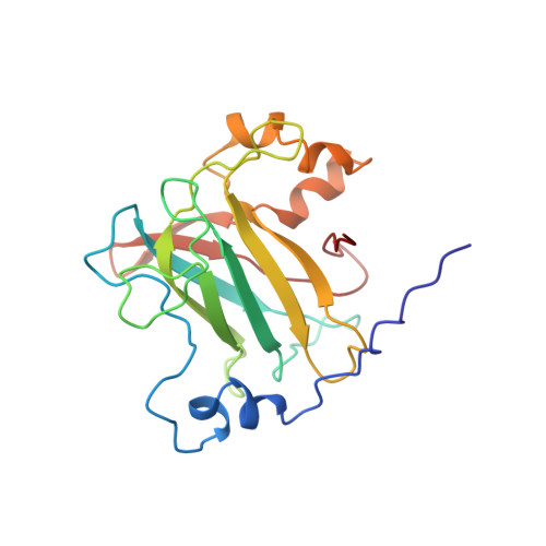
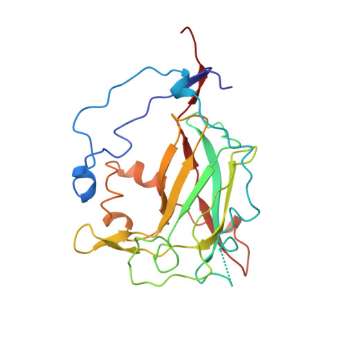
Structures of competitive inhibitor complexes of protocatechuate 3,4-dioxygenase: multiple exogenous ligand binding orientations within the active site.

Orville, A.M.Elango, N.Lipscomb, J.D.Ohlendorf, D.H.

(1997) Biochemistry 36: 10039-10051

  • DOI: https://doi.org/10.1021/bi970468n
  • Primary Citation of Related Structures:  
    3PCB, 3PCC, 3PCE, 3PCF, 3PCG, 3PCH, 3PCI

  • PubMed Abstract: 

    Protocatechuate 3,4-dioxygenase (3,4-PCD) catalyzes the oxidative ring cleavage of 3,4-dihydroxybenzoate to produce beta-carboxy-cis, cis-muconate. Crystal structures of Pseudomonas putida3,4-PCD [quaternary structure of (alphabetaFe3+)12] complexed with seven competitive inhibitors [3-hydroxyphenylacetate (MHP), 4-hydroxyphenylacetate (PHP), 3-hydroxybenzoate (MHB), 4-hydroxybenzoate (PHB), 3-fluoro-4-hydroxybenzoate (FHB), 3-chloro-4-hydroxybenzoate (CHB), and 3-iodo-4-hydroxybenzoate (IHB)] are reported at 2.0-2.2 A resolution with R-factors of 0. 0.159-0.179. The inhibitors bind in a narrow active site crevasse lined with residues that provide a microenvironment that closely matches the chemical characteristics of the inhibitors. This results in as little as 20% solvent-exposed surface area for the higher-affinity inhibitors (PHB, CHB, and FHB). In uncomplexed 3,4-PCD, the active site Fe3+ is bound at the bottom of the active site crevasse by four endogenous ligands and a solvent molecule (Wat827). The orientations of the endogenous ligands are relatively unperturbed in each inhibitor complex, but the inhibitors themselves bind to or near the iron in a range of positions, all of which perturb the position of Wat827. The three lowest-affinity inhibitors (MHP, PHP, and IHB) yield distorted trigonal bipyramidal iron coordination geometry in which the inhibitor C4-phenolate group displaces the solvent ligand. MHB binds within the active site, but neither its C3-OH group nor the solvent molecule binds to the iron. The C4-phenolate group of the three highest-affinity inhibitors (PHB, CHB, and FHB) coordinates the Fe3+ adjacent to Wat827, resulting in a shift in its position to yield a six-coordinate distorted octahedral geometry. The range of inhibitor orientations may mimic the mechanistically significant stages of substrate binding to 3, 4-PCD. The structure of the final substrate complex is reported in the following paper [Orville, A. M., Lipscomb, J. D., & Ohlendorf, D. H. (1997) Biochemistry 36, 10052-10066].


  • Organizational Affiliation

    Department of Biochemistry, Medical School, and Center for Metals in Biocatalysis, University of Minnesota, Minneapolis, Minnesota 55455-0347, USA.


Macromolecules
Find similar proteins by:  (by identity cutoff)  |  3D Structure
Entity ID: 1
MoleculeChains Sequence LengthOrganismDetailsImage
Protocatechuate 3,4-dioxygenase alpha chain200Pseudomonas putidaMutation(s): 0 
EC: 1.13.11.3
UniProt
Find proteins for P00436 (Pseudomonas putida)
Explore P00436 
Go to UniProtKB:  P00436
Entity Groups  
Sequence Clusters30% Identity50% Identity70% Identity90% Identity95% Identity100% Identity
UniProt GroupP00436
Sequence Annotations
Expand
  • Reference Sequence
Find similar proteins by:  (by identity cutoff)  |  3D Structure
Entity ID: 2
MoleculeChains Sequence LengthOrganismDetailsImage
Protocatechuate 3,4-dioxygenase beta chain238Pseudomonas putidaMutation(s): 0 
EC: 1.13.11.3
UniProt
Find proteins for P00437 (Pseudomonas putida)
Explore P00437 
Go to UniProtKB:  P00437
Entity Groups  
Sequence Clusters30% Identity50% Identity70% Identity90% Identity95% Identity100% Identity
UniProt GroupP00437
Sequence Annotations
Expand
  • Reference Sequence
Small Molecules
Ligands 2 Unique
IDChains Name / Formula / InChI Key2D Diagram3D Interactions
CHB
Query on CHB

Download Ideal Coordinates CCD File 
AA [auth Q]
CA [auth R]
DA [auth R]
N [auth M]
O [auth M]
AA [auth Q],
CA [auth R],
DA [auth R],
N [auth M],
O [auth M],
Q [auth N],
R [auth N],
T [auth O],
U [auth O],
W [auth P],
X [auth P],
Z [auth Q]
3-CHLORO-4-HYDROXYBENZOIC ACID
C7 H5 Cl O3
QGNLHMKIGMZKJX-UHFFFAOYSA-N
FE
Query on FE

Download Ideal Coordinates CCD File 
BA [auth R]
M
P [auth N]
S [auth O]
V [auth P]
BA [auth R],
M,
P [auth N],
S [auth O],
V [auth P],
Y [auth Q]
FE (III) ION
Fe
VTLYFUHAOXGGBS-UHFFFAOYSA-N
Modified Residues  1 Unique
IDChains TypeFormula2D DiagramParent
CME
Query on CME
B [auth M]
D [auth N]
F [auth O]
H [auth P]
J [auth Q]
B [auth M],
D [auth N],
F [auth O],
H [auth P],
J [auth Q],
L [auth R]
L-PEPTIDE LINKINGC5 H11 N O3 S2CYS
Experimental Data & Validation

Experimental Data

  • Method: X-RAY DIFFRACTION
  • Resolution: 2.05 Å
  • R-Value Work:  0.167 (Depositor) 
Space Group: I 1 2 1
Unit Cell:
Length ( Å )Angle ( ˚ )
a = 195.39α = 90
b = 126.52β = 97.77
c = 133.33γ = 90
Software Package:
Software NamePurpose
PROLSQrefinement
XENGENdata reduction
XENGENdata scaling

Structure Validation

View Full Validation Report


Entry History 

Deposition Data

Revision History  (Full details and data files)

  • Version 1.0: 1998-01-07
    Type: Initial release
  • Version 1.1: 2008-03-03
    Changes: Version format compliance
  • Version 1.2: 2011-07-13
    Changes: Advisory, Derived calculations, Version format compliance
  • Version 2.0: 2023-09-27
    Type: Remediation
    Changes: Advisory, Atomic model, Data collection, Database references, Derived calculations, Other, Refinement description
  • Version 3.0: 2024-12-25
    Changes: Advisory, Atomic model, Data collection, Database references, Derived calculations, Non-polymer description, Polymer sequence, Source and taxonomy, Structure summary