3P5A | pdb_00003p5a

Human Carbonic Anhydrase complexed with Sodium morpholinocarbodithioate


Experimental Data Snapshot

  • Method: X-RAY DIFFRACTION
  • Resolution: 1.49 Å
  • R-Value Free: 
    0.169 (Depositor), 0.170 (DCC) 
  • R-Value Work: 
    0.148 (Depositor), 0.149 (DCC) 
  • R-Value Observed: 
    0.149 (Depositor) 

wwPDB Validation 3D Report Full Report

Validation slider image for 3P5A

Ligand Structure Quality Assessment 


This is version 1.3 of the entry. See complete history

Literature

Dithiocarbamates strongly inhibit carbonic anhydrases and show antiglaucoma action in vivo

Carta, F.Aggarwal, M.Maresca, A.Scozzafava, A.McKenna, R.Masini, E.Supuran, C.T.

(2012) J Med Chem 55: 1721-1730

  • DOI: https://doi.org/10.1021/jm300031j
  • Primary Citation Related Structures: 
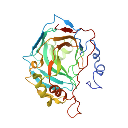
    3P5A

  • PubMed Abstract: 

    A series of dithiocarbamates were prepared by reaction of primary/secondary amines with carbon disulfide in the presence of bases. These compounds were tested for the inhibition of four human (h) isoforms of the zinc enzyme carbonic anhydrase, CA (EC 4.2.1.1), hCA I, II, IX, and XII, involved in pathologies such as glaucoma (CA II and XII) or cancer (CA IX). Several low nanomolar inhibitors targeting these CAs were detected. The X-ray crystal structure of the hCA II adduct with morpholine dithiocarbamate evidenced the inhibition mechanism of these compounds, which coordinate to the metal ion through a sulfur atom from the dithiocarbamate zinc-binding function. Some dithiocarbamates showed an effective intraocular pressure lowering activity in an animal model of glucoma.


  • Organizational Affiliation
    • Laboratorio di Chimica Bioinorganica, Polo Scientifico, Università degli Studi di Firenze, Room 188, Via della Lastruccia 3, 50019 Sesto Fiorentino, Florence, Italy.

Macromolecule Content 

  • Total Structure Weight: 29.6 kDa 
  • Atom Count: 2,392 
  • Modeled Residue Count: 257 
  • Deposited Residue Count: 260 
  • Unique protein chains: 1

Macromolecules

Find similar proteins by:|  3D Structure
Entity ID: 1
MoleculeChains  Sequence LengthOrganismDetailsImage
Carbonic anhydrase 2260Homo sapiensMutation(s): 0 
Gene Names: CA2
EC: 4.2.1.1 (PDB Primary Data), 4.2.1.69 (UniProt)
UniProt & NIH Common Fund Data Resources
Find proteins for P00918 (Homo sapiens)
Explore P00918 
Go to UniProtKB:  P00918
PHAROS:  P00918
GTEx:  ENSG00000104267 
Entity Groups
Sequence Clusters30% Identity50% Identity70% Identity90% Identity95% Identity100% Identity
UniProt GroupP00918
Sequence Annotations
Expand
Reference Sequence

Experimental Data & Validation

Experimental Data

  • Method: X-RAY DIFFRACTION
  • Resolution: 1.49 Å
  • R-Value Free:  0.169 (Depositor), 0.170 (DCC) 
  • R-Value Work:  0.148 (Depositor), 0.149 (DCC) 
  • R-Value Observed: 0.149 (Depositor) 
Space Group: P 1 21 1
Unit Cell:
Length ( Å )Angle ( ˚ )
a = 42.262α = 90
b = 41.21β = 104.21
c = 72.151γ = 90
Software Package:
Software NamePurpose
DENZOdata reduction
SCALEPACKdata scaling
PHENIXrefinement
PDB_EXTRACTdata extraction
CrystalCleardata collection
PHENIXphasing

Structure Validation

View Full Validation Report


Ligand Structure Quality Assessment 


Entry History 

Deposition Data

Revision History  (Full details and data files)

  • Version 1.0: 2011-10-19
    Type: Initial release
  • Version 1.1: 2012-01-25
    Changes: Database references
  • Version 1.2: 2017-11-08
    Changes: Refinement description
  • Version 1.3: 2024-02-21
    Changes: Data collection, Database references, Derived calculations