3P55 | pdb_00003p55

Human carbonic anhydrase II in complex with p-(4-ferrocenyl-1H-1,2,3-triazol-1-yl)benzenesulfonamide


Experimental Data Snapshot

  • Method: X-RAY DIFFRACTION
  • Resolution: 2.00 Å
  • R-Value Free: 
    0.223 (Depositor), 0.188 (DCC) 
  • R-Value Work: 
    0.187 (Depositor), 0.182 (DCC) 

Starting Model: experimental
View more details

wwPDB Validation 3D Report Full Report

Validation slider image for 3P55

Ligand Structure Quality Assessment 


This is version 1.3 of the entry. See complete history

Literature

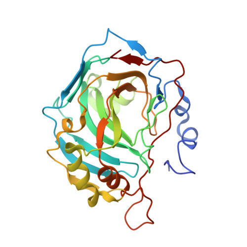
Protein crystal structures with ferrocene and ruthenocene-based enzyme inhibitors.

Salmon, A.J.Williams, M.L.Hofmann, A.Poulsen, S.A.

(2012) Chem Commun (Camb) 48: 2328-2330

  • DOI: https://doi.org/10.1039/c2cc15625c
  • Primary Citation Related Structures: 
    3P3H, 3P3J, 3P44, 3P55

  • PubMed Abstract: 

    We have determined the protein X-ray crystal structures of four organometallic inhibitors in complex with their target enzyme carbonic anhydrase II. The barrel-shaped hydrophobic ferrocene and ruthenocene moieties have provided a structure-based avenue to better occupy the hydrophobic binding patch within the enzyme active site.


  • Organizational Affiliation
    • Eskitis Institute for Cell and Molecular Therapies, Griffith University, Nathan, Queensland 4111, Australia.

Macromolecule Content 

  • Total Structure Weight: 29.94 kDa 
  • Atom Count: 2,260 
  • Modeled Residue Count: 257 
  • Deposited Residue Count: 260 
  • Unique protein chains: 1

Macromolecules

Find similar proteins by:|  3D Structure
Entity ID: 1
MoleculeChains  Sequence LengthOrganismDetailsImage
Carbonic anhydrase 2260Homo sapiensMutation(s): 0 
Gene Names: CA2
EC: 4.2.1.1 (PDB Primary Data), 4.2.1.69 (UniProt)
UniProt & NIH Common Fund Data Resources
Find proteins for P00918 (Homo sapiens)
Explore P00918 
Go to UniProtKB:  P00918
PHAROS:  P00918
GTEx:  ENSG00000104267 
Entity Groups
Sequence Clusters30% Identity50% Identity70% Identity90% Identity95% Identity100% Identity
UniProt GroupP00918
Sequence Annotations
Expand
Reference Sequence

Experimental Data & Validation

Experimental Data

  • Method: X-RAY DIFFRACTION
  • Resolution: 2.00 Å
  • R-Value Free:  0.223 (Depositor), 0.188 (DCC) 
  • R-Value Work:  0.187 (Depositor), 0.182 (DCC) 
Space Group: P 1 21 1
Unit Cell:
Length ( Å )Angle ( ˚ )
a = 42.4α = 90
b = 41.6β = 104.64
c = 72.23γ = 90
Software Package:
Software NamePurpose
Blu-Icedata collection
CNSrefinement
MOSFLMdata reduction
SCALAdata scaling

Structure Validation

View Full Validation Report


Ligand Structure Quality Assessment 


Entry History 

Deposition Data

  • Released Date: 2011-10-12 
  • Deposition Author(s): Salmon, A.J.

Revision History  (Full details and data files)

  • Version 1.0: 2011-10-12
    Type: Initial release
  • Version 1.1: 2012-02-08
    Changes: Database references
  • Version 1.2: 2013-08-28
    Changes: Database references
  • Version 1.3: 2023-11-01
    Changes: Data collection, Database references, Derived calculations, Refinement description