3OWN | pdb_00003own

Potent macrocyclic renin inhibitors


Experimental Data Snapshot

  • Method: X-RAY DIFFRACTION
  • Resolution: 2.00 Å
  • R-Value Free: 
    0.254 (Depositor), 0.253 (DCC) 
  • R-Value Work: 
    0.217 (Depositor), 0.215 (DCC) 
  • R-Value Observed: 
    0.219 (Depositor) 

wwPDB Validation 3D Report Full Report

Validation slider image for 3OWN

Ligand Structure Quality Assessment 


This is version 1.2 of the entry. See complete history

Literature

Design and synthesis of potent macrocyclic renin inhibitors.

Sund, C.Belda, O.Wiktelius, D.Sahlberg, C.Vrang, L.Sedig, S.Hamelink, E.Henderson, I.Agback, T.Jansson, K.Borkakoti, N.Derbyshire, D.Eneroth, A.Samuelsson, B.

(2011) Bioorg Med Chem Lett 21: 358-362

  • DOI: https://doi.org/10.1016/j.bmcl.2010.10.140
  • Primary Citation Related Structures: 
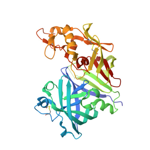
    3OWN

  • PubMed Abstract: 

    Two types of P1-P3-linked macrocyclic renin inhibitors containing the hydroxyethylene isostere (HE) scaffold just outside the macrocyclic ring have been synthesized. An aromatic or aliphatic substituent (P3sp) was introduced in the macrocyclic ring aiming at the S3 subpocket (S3sp) in order to optimize the potency. A 5-6-fold improvement in both the K(i) and the human plasma renin activity (HPRA)IC(50) was observed when moving from the starting linear peptidomimetic compound 1 to the most potent macrocycle 42 (K(i) = 3.3 nM and HPRA IC(50) = 7 nM). Truncation of the prime side of 42 led to 8-10-fold loss of inhibitory activity in macrocycle 43 (K(i) = 34 nM and HPRA IC(50) = 56 nM). All macrocycles were epimeric mixtures in regard to the P3sp substituent and X-ray crystallographic data of the representative renin macrocycle 43 complex showed that only the S-isomer buried the substituent into the S3sp. Inhibitory selectivity over cathepsin D (Cat-D) and BACE-1 was also investigated for all the macrocycles and showed that truncation of the prime side increased selectivity of inhibition in favor of renin.


  • Organizational Affiliation
    • Medivir AB, PO Box 1086, SE-14122 Huddinge, Sweden. christian.sund@medivir.se

Macromolecule Content 

  • Total Structure Weight: 76.42 kDa 
  • Atom Count: 5,603 
  • Modeled Residue Count: 668 
  • Deposited Residue Count: 682 
  • Unique protein chains: 1

Macromolecules

Find similar proteins by:|  3D Structure
Entity ID: 1
MoleculeChains  Sequence LengthOrganismDetailsImage
Renin
A, B
341Homo sapiensMutation(s): 0 
EC: 3.4.23.15
UniProt & NIH Common Fund Data Resources
Find proteins for P00797 (Homo sapiens)
Explore P00797 
Go to UniProtKB:  P00797
PHAROS:  P00797
GTEx:  ENSG00000143839 
Entity Groups
Sequence Clusters30% Identity50% Identity70% Identity90% Identity95% Identity100% Identity
UniProt GroupP00797
Sequence Annotations
Expand
Reference Sequence

Small Molecules

Ligands 4 Unique
IDChains Name / Formula / InChI Key2D Diagram3D Interactions
3OX

Query on 3OX


Download:Ideal Coordinates CCD File
H [auth A](2S,4S)-4-hydroxy-2-(1-methylethyl)-4-[(4R,13S)-18-[methyl(methylsulfonyl)amino]-2,15-dioxo-4-phenyl-11-oxa-3,14-diazatricyclo[14.3.1.1~5,9~]henicosa-1(20),5(21),6,8,16,18-hexaen-13-yl]-N-(2-methylpropyl)butanamide
C37 H48 N4 O7 S
NUOROLSRIMMSEX-GZXHTMMISA-N
3OW

Query on 3OW


Download:Ideal Coordinates CCD File
I [auth B](2S,4S)-4-hydroxy-2-(1-methylethyl)-4-[(4S,13S)-18-[methyl(methylsulfonyl)amino]-2,15-dioxo-4-phenyl-11-oxa-3,14-diazatricyclo[14.3.1.1~5,9~]henicosa-1(20),5(21),6,8,16,18-hexaen-13-yl]-N-(2-methylpropyl)butanamide
C37 H48 N4 O7 S
NUOROLSRIMMSEX-CUPIEXAXSA-N
ACT

Query on ACT


Download:Ideal Coordinates CCD File
C [auth A],
D [auth A],
G [auth A]
ACETATE ION
C2 H3 O2
QTBSBXVTEAMEQO-UHFFFAOYSA-M
NA

Query on NA


Download:Ideal Coordinates CCD File
E [auth A],
F [auth A]
SODIUM ION
Na
FKNQFGJONOIPTF-UHFFFAOYSA-N

Experimental Data & Validation

Experimental Data

  • Method: X-RAY DIFFRACTION
  • Resolution: 2.00 Å
  • R-Value Free:  0.254 (Depositor), 0.253 (DCC) 
  • R-Value Work:  0.217 (Depositor), 0.215 (DCC) 
  • R-Value Observed: 0.219 (Depositor) 
Space Group: P 21 3
Unit Cell:
Length ( Å )Angle ( ˚ )
a = 141.827α = 90
b = 141.827β = 90
c = 141.827γ = 90
Software Package:
Software NamePurpose
MOSFLMdata reduction
SCALAdata scaling
PHASERphasing
REFMACrefinement
PDB_EXTRACTdata extraction

Structure Validation

View Full Validation Report


Ligand Structure Quality Assessment 


Entry History 

Deposition Data

Revision History  (Full details and data files)

  • Version 1.0: 2010-12-15
    Type: Initial release
  • Version 1.1: 2011-07-13
    Changes: Version format compliance
  • Version 1.2: 2024-11-20
    Changes: Data collection, Database references, Derived calculations, Structure summary