


The crystal structure of an amidohydrolase from Mycoplasma synoviae with Zn ion bound
Zhang, Z., Kumaran, D., Burley, S.K., Swaminathan, S.To be published.
Experimental Data Snapshot
Starting Model: experimental
View more details
wwPDB Validation 3D Report Full Report

Macromolecule Content
Entity ID: 1 | |||||
|---|---|---|---|---|---|
| Molecule | Chains | Sequence Length | Organism | Details | Image |
| amidohydrolase | 363 | Mycoplasmopsis synoviae 53 | Mutation(s): 0 Gene Names: had, MS53_0025 EC: 3.1.1.104 (UniProt), 3.1.1.25 (UniProt) | 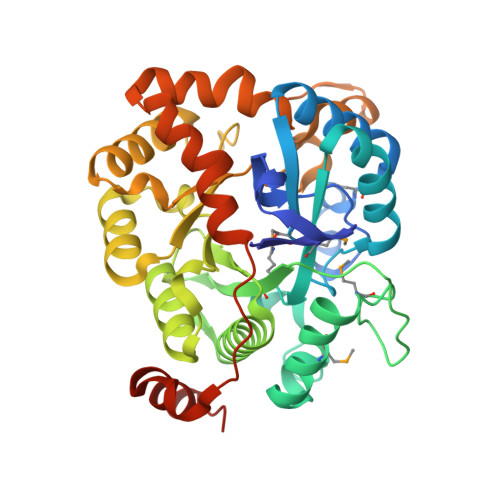 | |
UniProt | |||||
Entity Groups | |||||
| Sequence Clusters | 30% Identity50% Identity70% Identity90% Identity95% Identity100% Identity | ||||
| UniProt Group | Q4A724 | ||||
Sequence AnnotationsExpand | |||||
Reference Sequence | |||||
| Ligands 2 Unique | |||||
|---|---|---|---|---|---|
| ID | Chains | Name / Formula / InChI Key | 2D Diagram | 3D Interactions | |
| PO4 Download:Ideal Coordinates CCD File | I [auth A] L [auth B] O [auth C] R [auth D] U [auth E] | PHOSPHATE ION O4 P NBIIXXVUZAFLBC-UHFFFAOYSA-K |  | ||
| ZN Download:Ideal Coordinates CCD File | G [auth A] H [auth A] J [auth B] K [auth B] M [auth C] | ZINC ION Zn PTFCDOFLOPIGGS-UHFFFAOYSA-N |  | ||
| Modified Residues 2 Unique | |||||
|---|---|---|---|---|---|
| ID | Chains | Type | Formula | 2D Diagram | Parent |
| KCX Query on KCX | A, B, C, D, E A, B, C, D, E, F | L-PEPTIDE LINKING | C7 H14 N2 O4 |  | LYS |
| MSE Query on MSE | A, B, C, D, E A, B, C, D, E, F | L-PEPTIDE LINKING | C5 H11 N O2 Se |  | MET |
| Length ( Å ) | Angle ( ˚ ) |
|---|---|
| a = 89.256 | α = 98.95 |
| b = 89.186 | β = 92.89 |
| c = 96.059 | γ = 119.86 |
| Software Name | Purpose |
|---|---|
| CBASS | data collection |
| PHASER | phasing |
| PHENIX | refinement |
| HKL-2000 | data reduction |
| HKL-2000 | data scaling |


