3OUX | pdb_00003oux

Structure of beta-catenin with phosphorylated Lef-1


Experimental Data Snapshot

  • Method: X-RAY DIFFRACTION
  • Resolution: 2.40 Å
  • R-Value Free: 
    0.264 (Depositor), 0.265 (DCC) 
  • R-Value Work: 
    0.199 (Depositor), 0.198 (DCC) 

Starting Model: experimental
View more details

wwPDB Validation 3D Report Full Report

Validation slider image for 3OUX

This is version 1.5 of the entry. See complete history

Literature

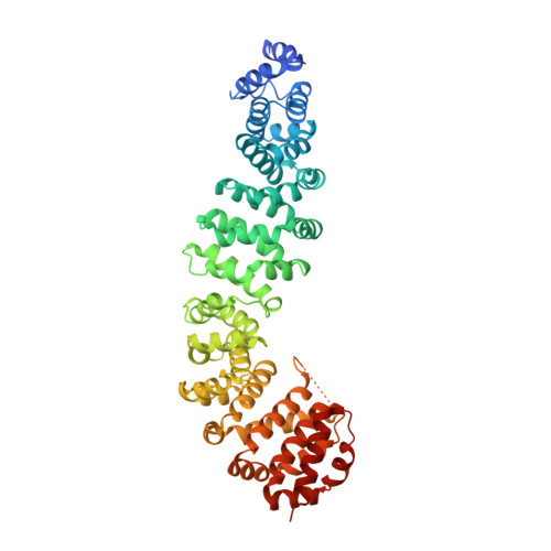
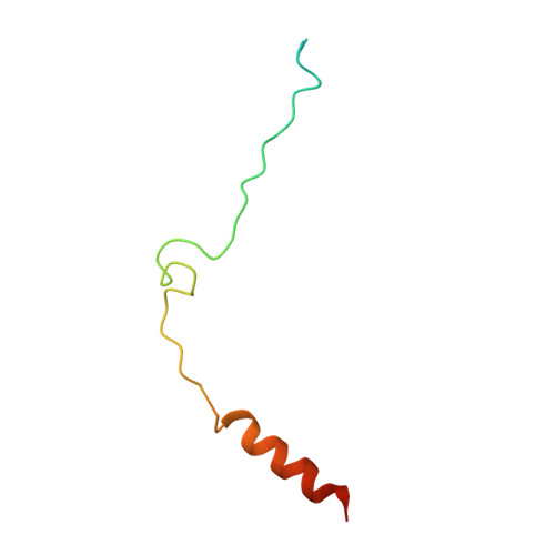
Biochemical and structural characterization of beta-catenin interactions with nonphosphorylated and CK2-phosphorylated Lef-1.

Sun, J.Weis, W.I.

(2011) J Mol Biology 405: 519-530

  • DOI: https://doi.org/10.1016/j.jmb.2010.11.010
  • Primary Citation Related Structures: 
    3OUW, 3OUX

  • PubMed Abstract: 

    In the Wnt/β-catenin signaling pathway, β-catenin activates target genes through its interactions with the T-cell factor/lymphoid enhancer-binding factor (TCF/Lef) family of transcription factors. The crystal structures of complexes between the β-catenin armadillo domain and the Lef-1 N-terminal domain show that the overall conformation and many of the interactions are similar to other published structures of TCFs bound to β-catenin. However, a second salt bridge in other TCF-β-catenin structures is absent in the structure of β-catenin-Lef-1 complex, indicating that this feature is not obligatory for β-catenin binding. Casein kinase II (CK2) has been shown to act as a positive regulator of Wnt signaling, and Lef-1 is a substrate of CK2. In vitro phosphorylation of purified Lef-1 was used to examine the effect of CK2 on the interaction of Lef-1 with β-catenin. Mass spectrometry data show that CK2 phosphorylation of Lef-1 N-terminal domain results in a single phosphorylation site at Ser40. Isothermal titration calorimetry revealed that β-catenin binds to nonphosphorylated or CK2-phosphorylated Lef-1 with the same affinity, which is consistent with the absence of phospho-Ser40 interactions in the crystal structure of phosphorylated Lef-1 N-terminal domain bound to β-catenin. These data indicate that the effect of CK2 on the Wnt/β-catenin pathway does not appear to be at the level of the Lef-1-β-catenin interaction.


  • Organizational Affiliation
    • Department of Structural Biology, Stanford University School of Medicine,Stanford, CA 94305-5126, USA.

Macromolecule Content 

  • Total Structure Weight: 66.01 kDa 
  • Atom Count: 4,164 
  • Modeled Residue Count: 550 
  • Deposited Residue Count: 607 
  • Unique protein chains: 2

Macromolecules

Find similar proteins by:|  3D Structure
Entity ID: 1
MoleculeChains  Sequence LengthOrganismDetailsImage
Catenin beta-1540Mus musculusMutation(s): 0 
Gene Names: Ctnnb1Catnb
UniProt & NIH Common Fund Data Resources
Find proteins for Q02248 (Mus musculus)
Explore Q02248 
Go to UniProtKB:  Q02248
IMPC:  MGI:88276
Entity Groups
Sequence Clusters30% Identity50% Identity70% Identity90% Identity95% Identity100% Identity
UniProt GroupQ02248
Sequence Annotations
Expand
Reference Sequence
Find similar proteins by:|  3D Structure
Entity ID: 2
MoleculeChains  Sequence LengthOrganismDetailsImage
Lymphoid enhancer-binding factor 167Mus musculusMutation(s): 0 
Gene Names: Lef1Lef-1
UniProt & NIH Common Fund Data Resources
Find proteins for P27782 (Mus musculus)
Explore P27782 
Go to UniProtKB:  P27782
IMPC:  MGI:96770
Entity Groups
Sequence Clusters30% Identity50% Identity70% Identity90% Identity95% Identity100% Identity
UniProt GroupP27782
Sequence Annotations
Expand
Reference Sequence

Experimental Data & Validation

Experimental Data

  • Method: X-RAY DIFFRACTION
  • Resolution: 2.40 Å
  • R-Value Free:  0.264 (Depositor), 0.265 (DCC) 
  • R-Value Work:  0.199 (Depositor), 0.198 (DCC) 
Space Group: I 4 2 2
Unit Cell:
Length ( Å )Angle ( ˚ )
a = 115.56α = 90
b = 115.56β = 90
c = 195.93γ = 90
Software Package:
Software NamePurpose
Blu-Icedata collection
PHASERphasing
PHENIXrefinement
MOSFLMdata reduction
SCALAdata scaling

Structure Validation

View Full Validation Report


Entry History 

Deposition Data

Revision History  (Full details and data files)

  • Version 1.0: 2010-11-24
    Type: Initial release
  • Version 1.1: 2011-07-13
    Changes: Version format compliance
  • Version 1.2: 2015-12-02
    Changes: Database references
  • Version 1.3: 2015-12-09
    Changes: Other
  • Version 1.4: 2023-09-06
    Changes: Data collection, Database references, Refinement description
  • Version 1.5: 2024-10-30
    Changes: Structure summary