3OFW

Crystal structure of recombinant Kunitz Type serine protease Inhibitor-1 from the Carribean sea anemone stichodactyla helianthus


Experimental Data Snapshot

  • Method: X-RAY DIFFRACTION
  • Resolution: 2.50 Å
  • R-Value Free: 0.221 
  • R-Value Work: 0.190 
  • R-Value Observed: 0.191 

wwPDB Validation   3D Report Full Report


This is version 1.2 of the entry. See complete history


Literature

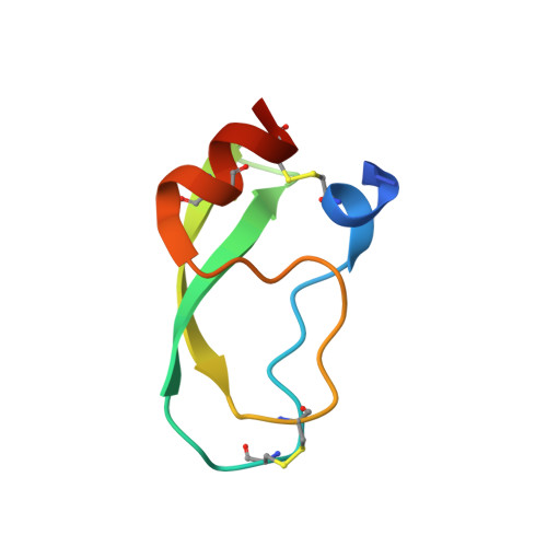
Structure of the recombinant BPTI/Kunitz-type inhibitor rShPI-1A from the marine invertebrate Stichodactyla helianthus.

Garcia-Fernandez, R.Pons, T.Meyer, A.Perbandt, M.Gonzalez-Gonzalez, Y.Gil, D.de los Angeles Chavez, M.Betzel, C.Redecke, L.

(2012) Acta Crystallogr Sect F Struct Biol Cryst Commun 68: 1289-1293

  • DOI: 10.1107/S1744309112039085
  • Primary Citation of Related Structures:  
    3OFW

  • PubMed Abstract: 
  • The BPTI/Kunitz-type inhibitor family includes several extremely potent serine protease inhibitors. To date, the inhibitory mechanisms have only been studied for mammalian inhibitors. Here, the first crystal structure of a BPTI/Kunitz-type inhibitor from a marine invertebrate (rShPI-1A) is reported to 2 ...

    The BPTI/Kunitz-type inhibitor family includes several extremely potent serine protease inhibitors. To date, the inhibitory mechanisms have only been studied for mammalian inhibitors. Here, the first crystal structure of a BPTI/Kunitz-type inhibitor from a marine invertebrate (rShPI-1A) is reported to 2.5 Å resolution. Crystallization of recombinant rShPI-1A required the salt-induced dissociation of a trypsin complex that was previously formed to avoid intrinsic inhibitor aggregates in solution. The rShPI-1A structure is similar to the NMR structure of the molecule purified from the natural source, but allowed the assignment of disulfide-bridge chiralities and the detection of an internal stabilizing water network. A structural comparison with other BPTI/Kunitz-type canonical inhibitors revealed unusual ϕ angles at positions 17 and 30 to be a particular characteristic of the family. A significant clustering of ϕ and ψ angle values in the glycine-rich remote fragment near the secondary binding loop was additionally identified, but its impact on the specificity of rShPI-1A and similar molecules requires further study.


    Organizational Affiliation

    Centro de Estudio de Proteínas, Facultad de Biología, Universidad de la Habana, Calle 25 No. 455, Havana 10400, Cuba.


Macromolecules
Find similar proteins by:  (by identity cutoff)  |  3D Structure
Entity ID: 1
MoleculeChainsSequence LengthOrganismDetailsImage
Kunitz-type proteinase inhibitor SHPI-1A60Stichodactyla helianthusMutation(s): 0 
UniProt
Find proteins for P31713 (Stichodactyla helianthus)
Explore P31713 
Go to UniProtKB:  P31713
Entity Groups  
Sequence Clusters30% Identity50% Identity70% Identity90% Identity95% Identity100% Identity
UniProt GroupP31713
Protein Feature View
Expand
  • Reference Sequence
Small Molecules
Ligands 1 Unique
IDChainsName / Formula / InChI Key2D Diagram3D Interactions
CL
Query on CL

Download Ideal Coordinates CCD File 
B [auth A],
C [auth A],
D [auth A]
CHLORIDE ION
Cl
VEXZGXHMUGYJMC-UHFFFAOYSA-M
 Ligand Interaction
Experimental Data & Validation

Experimental Data

  • Method: X-RAY DIFFRACTION
  • Resolution: 2.50 Å
  • R-Value Free: 0.221 
  • R-Value Work: 0.190 
  • R-Value Observed: 0.191 
  • Space Group: P 43 21 2
Unit Cell:
Length ( Å )Angle ( ˚ )
a = 37.158α = 90
b = 37.158β = 90
c = 114.98γ = 90
Software Package:
Software NamePurpose
MAR345dtbdata collection
AMoREphasing
REFMACrefinement
DENZOdata reduction
SCALAdata scaling

Structure Validation

View Full Validation Report


Entry History 

Deposition Data

Revision History  (Full details and data files)

  • Version 1.0: 2011-08-17
    Type: Initial release
  • Version 1.1: 2012-11-07
    Changes: Database references
  • Version 1.2: 2013-01-23
    Changes: Database references