Total chemical synthesis and X-ray structure of kaliotoxin by racemic protein crystallography.
Pentelute, B.L., Mandal, K., Gates, Z.P., Sawaya, M.R., Yeates, T.O., Kent, S.B.(2010) Chem Commun (Camb) 46: 8174-8176
- PubMed: 20877851 Search on PubMed
- DOI: https://doi.org/10.1039/c0cc03148h
- Primary Citation Related Structures:
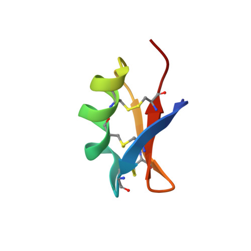
3ODV - PubMed Abstract:
Here we report the total synthesis of kaliotoxin by 'one pot' native chemical ligation of three synthetic peptides. A racemic mixture of D- and L-kaliotoxin synthetic protein molecules gave crystals in the centrosymmetric space group P1 that diffracted to atomic-resolution (0.95 Å), enabling the X-ray structure of kaliotoxin to be determined by direct methods.
- Department of Chemistry, Institute for Biophysical Dynamics, The University of Chicago, Chicago, Illinois 60637, USA.
Organizational Affiliation: