3OBU

Crystal structure of the Tsg101 UEV domain in complex with a HIV-1 PTAP peptide


Experimental Data Snapshot

  • Method: X-RAY DIFFRACTION
  • Resolution: 1.60 Å
  • R-Value Free: 0.236 
  • R-Value Work: 0.199 
  • R-Value Observed: 0.199 

wwPDB Validation   3D Report Full Report


This is version 1.1 of the entry. See complete history


Literature

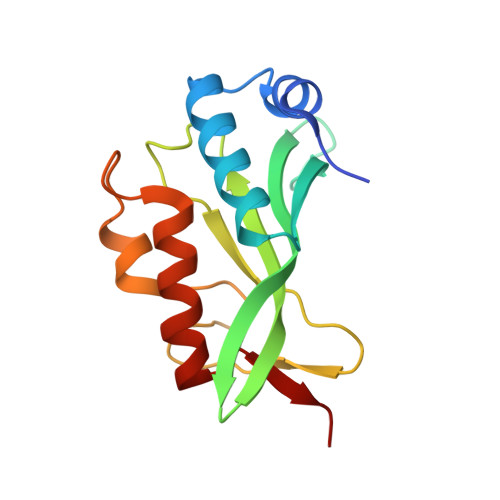
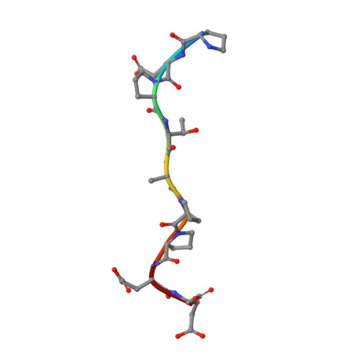
Crystallographic and Functional Analysis of the ESCRT-I /HIV-1 Gag PTAP Interaction.

Im, Y.J.Kuo, L.Ren, X.Burgos, P.V.Zhao, X.Z.Liu, F.Burke, T.R.Bonifacino, J.S.Freed, E.O.Hurley, J.H.

(2010) Structure 18: 1536-1547

  • DOI: https://doi.org/10.1016/j.str.2010.08.010
  • Primary Citation of Related Structures:  
    3OBQ, 3OBS, 3OBU, 3OBX

  • PubMed Abstract: 
  • Budding of HIV-1 requires the binding of the PTAP late domain of the Gag p6 protein to the UEV domain of the TSG101 subunit of ESCRT-I. The normal function of this motif in cells is in receptor downregulation. Here, we report the 1.4-1.6 Å structures of the human TSG101 UEV domain alone and with wild-type and mutant HIV-1 PTAP and Hrs PSAP nonapeptides ...

    Budding of HIV-1 requires the binding of the PTAP late domain of the Gag p6 protein to the UEV domain of the TSG101 subunit of ESCRT-I. The normal function of this motif in cells is in receptor downregulation. Here, we report the 1.4-1.6 Å structures of the human TSG101 UEV domain alone and with wild-type and mutant HIV-1 PTAP and Hrs PSAP nonapeptides. The hydroxyl of the Thr or Ser residue in the P(S/T)AP motif hydrogen bonds with the main chain of Asn69. Mutation of the Asn to Pro, blocking the main-chain amide, abrogates PTAP motif binding in vitro and blocks budding of HIV-1 from cells. N69P and other PTAP binding-deficient alleles of TSG101 did not rescue HIV-1 budding. However, the mutant alleles did rescue downregulation of endogenous EGF receptor. This demonstrates that the PSAP motif is not rate determining in EGF receptor downregulation under normal conditions.


    Organizational Affiliation

    Laboratory of Molecular Biology, National Institute of Diabetes and Digestive and Kidney Diseases, National Institutes of Health, Bethesda, MD 20892-0580, USA.


Macromolecules
Find similar proteins by:  (by identity cutoff)  |  3D Structure
Entity ID: 1
MoleculeChainsSequence LengthOrganismDetailsImage
Tumor susceptibility gene 101 proteinA146Homo sapiensMutation(s): 0 
Gene Names: TSG101
UniProt & NIH Common Fund Data Resources
Find proteins for Q99816 (Homo sapiens)
Explore Q99816 
Go to UniProtKB:  Q99816
PHAROS:  Q99816
Entity Groups  
Sequence Clusters30% Identity50% Identity70% Identity90% Identity95% Identity100% Identity
UniProt GroupQ99816
Protein Feature View
Expand
  • Reference Sequence

Find similar proteins by:  Sequence   |   3D Structure  

Entity ID: 2
MoleculeChainsSequence LengthOrganismDetailsImage
Gag polyproteinB9N/AMutation(s): 0 
UniProt
Find proteins for Q72497 (Human immunodeficiency virus 1)
Explore Q72497 
Go to UniProtKB:  Q72497
Entity Groups  
UniProt GroupQ72497
Protein Feature View
Expand
  • Reference Sequence
Experimental Data & Validation

Experimental Data

  • Method: X-RAY DIFFRACTION
  • Resolution: 1.60 Å
  • R-Value Free: 0.236 
  • R-Value Work: 0.199 
  • R-Value Observed: 0.199 
  • Space Group: P 21 21 21
Unit Cell:
Length ( Å )Angle ( ˚ )
a = 33.526α = 90
b = 45.769β = 90
c = 85.446γ = 90
Software Package:
Software NamePurpose
HKL-2000data collection
MOLREPphasing
CNSrefinement
HKL-2000data reduction
HKL-2000data scaling

Structure Validation

View Full Validation Report


Entry History 

Deposition Data

Revision History  (Full details and data files)

  • Version 1.0: 2010-12-01
    Type: Initial release
  • Version 1.1: 2011-07-13
    Changes: Version format compliance