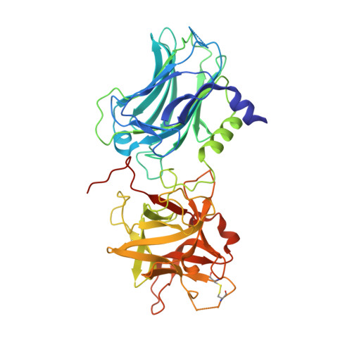
3OBT

Crystal structure of Botulinum neurotoxin serotype D ligand binding domain in complex with N-Acetylneuraminic acid


Experimental Data Snapshot

  • Method: X-RAY DIFFRACTION
  • Resolution: 2.00 Å
  • R-Value Free: 0.221 
  • R-Value Work: 0.172 
  • R-Value Observed: 0.174 

wwPDB Validation   3D Report Full Report


Ligand Structure Quality Assessment 


This is version 1.4 of the entry. See complete history


Literature

Botulinum neurotoxin serotype D attacks neurons via two carbohydrate-binding sites in a ganglioside-dependent manner.

Strotmeier, J.Lee, K.Volker, A.K.Mahrhold, S.Zong, Y.Zeiser, J.Zhou, J.Pich, A.Bigalke, H.Binz, T.Rummel, A.Jin, R.

(2010) Biochem J 431: 207-216

  • DOI: https://doi.org/10.1042/BJ20101042
  • Primary Citation of Related Structures:  
    3OBR, 3OBT

  • PubMed Abstract: 

    The extraordinarily high toxicity of botulinum neurotoxins primarily results from their specific binding and uptake into neurons. At motor neurons, the seven BoNT (botulinum neurotoxin) serotypes A-G inhibit acetylcholine release leading to flaccid paralysis. Uptake of BoNT/A, B, E, F and G requires a dual interaction with gangliosides and the synaptic vesicle proteins synaptotagmin or SV2 (synaptic vesicle glycoprotein 2), whereas little is known about the cell entry mechanisms of the serotypes C and D, which display the lowest amino acid sequence identity compared with the other five serotypes. In the present study we demonstrate that the neurotoxicity of BoNT/D depends on the presence of gangliosides by employing phrenic nerve hemidiaphragm preparations derived from mice expressing the gangliosides GM3, GM2, GM1 and GD1a, or only GM3 [a description of our use of ganglioside nomenclature is given in Svennerholm (1994) Prog. Brain Res. 101, XI-XIV]. High-resolution crystal structures of the 50 kDa cell-binding domain of BoNT/D alone and in complex with sialic acid, as well as biological analyses of single-site BoNT/D mutants identified two carbohydrate-binding sites. One site is located at a position previously identified in BoNT/A, B, E, F and G, but is lacking the conserved SXWY motif. The other site, co-ordinating one molecule of sialic acid, resembles the second ganglioside-binding pocket (the sialic-acid-binding site) of TeNT (tetanus neurotoxin).


  • Organizational Affiliation

    Institut für Toxikologie, Medizinische Hochschule Hannover, Carl-Neuberg-Strasse 1, Hannover, Germany.


Macromolecules
Find similar proteins by:  (by identity cutoff)  |  3D Structure
Entity ID: 1
MoleculeChains Sequence LengthOrganismDetailsImage
Botulinum neurotoxin type D434Clostridium botulinumMutation(s): 0 
Gene Names: botD
EC: 3.4.24.69
UniProt
Find proteins for P19321 (Clostridium botulinum D phage)
Explore P19321 
Go to UniProtKB:  P19321
Entity Groups  
Sequence Clusters30% Identity50% Identity70% Identity90% Identity95% Identity100% Identity
UniProt GroupP19321
Sequence Annotations
Expand
  • Reference Sequence
Small Molecules
Ligands 2 Unique
IDChains Name / Formula / InChI Key2D Diagram3D Interactions
SLB
Query on SLB

Download Ideal Coordinates CCD File 
B [auth A]N-acetyl-beta-neuraminic acid
C11 H19 N O9
SQVRNKJHWKZAKO-PFQGKNLYSA-N
GOL
Query on GOL

Download Ideal Coordinates CCD File 
C [auth A]
D [auth A]
E [auth A]
F [auth A]
G [auth A]
C [auth A],
D [auth A],
E [auth A],
F [auth A],
G [auth A],
H [auth A],
I [auth A],
J [auth A],
K [auth A],
L [auth A],
M [auth A]
GLYCEROL
C3 H8 O3
PEDCQBHIVMGVHV-UHFFFAOYSA-N
Experimental Data & Validation

Experimental Data

  • Method: X-RAY DIFFRACTION
  • Resolution: 2.00 Å
  • R-Value Free: 0.221 
  • R-Value Work: 0.172 
  • R-Value Observed: 0.174 
  • Space Group: P 21 21 21
Unit Cell:
Length ( Å )Angle ( ˚ )
a = 60.232α = 90
b = 90.044β = 90
c = 93.518γ = 90
Software Package:
Software NamePurpose
SCALEPACKdata scaling
REFMACrefinement
PDB_EXTRACTdata extraction
Blu-Icedata collection
DENZOdata reduction
PHASERphasing

Structure Validation

View Full Validation Report


Ligand Structure Quality Assessment 


Entry History 

Deposition Data

Revision History  (Full details and data files)

  • Version 1.0: 2010-09-08
    Type: Initial release
  • Version 1.1: 2011-07-13
    Changes: Version format compliance
  • Version 1.2: 2014-04-16
    Changes: Other
  • Version 1.3: 2017-11-08
    Changes: Refinement description
  • Version 1.4: 2020-07-29
    Type: Remediation
    Reason: Carbohydrate remediation
    Changes: Data collection, Database references, Derived calculations, Structure summary