Structural and Functional Studies of WlbA: A Dehydrogenase Involved in the Biosynthesis of 2,3-Diacetamido-2,3-dideoxy-d-mannuronic Acid .
Thoden, J.B., Holden, H.M.(2010) Biochemistry 49: 7939-7948
- PubMed: 20690587
- DOI: https://doi.org/10.1021/bi101103s
- Primary Citation of Related Structures:
3O9Z, 3OA0, 3OA2 - PubMed Abstract:
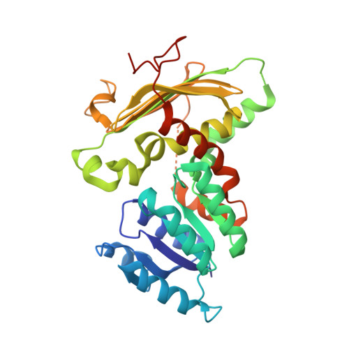
2,3-Diacetamido-2,3-dideoxy-d-mannuronic acid (ManNAc3NAcA) is an unusual dideoxy sugar first identified nearly 30 years ago in the lipopolysaccharide of Pseudomonas aeruginosa O:3a,d. It has since been observed in other organisms, including Bordetella pertussis, the causative agent of whooping cough. Five enzymes are required for the biosynthesis of UDP-ManNAc3NAcA starting from UDP-N-acetyl-d-glucosamine. Here we describe a structural study of WlbA, the NAD-dependent dehydrogenase that catalyzes the second step in the pathway, namely, the oxidation of the C-3' hydroxyl group on the UDP-linked sugar to a keto moiety and the reduction of NAD(+) to NADH. This enzyme has been shown to use alpha-ketoglutarate as an oxidant to regenerate the oxidized dinucleotide. For this investigation, three different crystal structures were determined: the enzyme with bound NAD(H), the enzyme in a complex with NAD(H) and alpha-ketoglutarate, and the enzyme in a complex with NAD(H) and its substrate (UDP-N-acetyl-d-glucosaminuronic acid). The tetrameric enzyme assumes an unusual quaternary structure with the dinucleotides positioned quite closely to one another. Both alpha-ketoglutarate and the UDP-linked sugar bind in the WlbA active site with their carbon atoms (C-2 and C-3', respectively) abutting the re face of the cofactor. They are positioned approximately 3 A from the nicotinamide C-4. The UDP-linked sugar substrate adopts a highly unusual curved conformation when bound in the WlbA active site cleft. Lys 101 and His 185 most likely play key roles in catalysis.
- Department of Biochemistry, University of Wisconsin, Madison, Wisconsin 53706, USA.
Organizational Affiliation:


