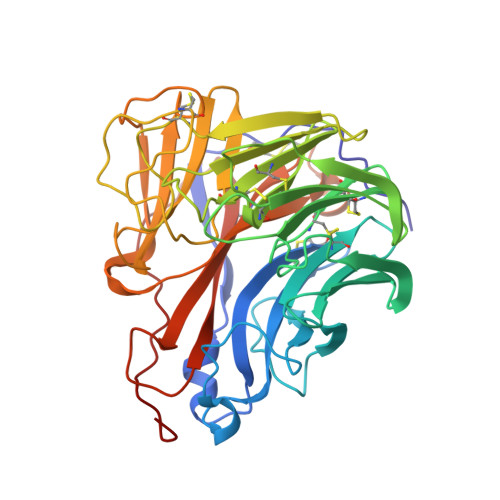
The 2009 pandemic H1N1 neuraminidase N1 lacks the 150-cavity in its active site
Li, Q., Qi, J.X., Zhang, W., Vavricka, C.J., Shi, Y., Wei, J.H., Feng, E.G., Shen, J.S., Chen, J.L., Liu, D., He, J.H., Yan, J.H., Liu, H., Jiang, H.L., Teng, M.K., Li, X.B., Gao, G.F.(2010) Nat Struct Mol Biol 17: 1266-1268
- PubMed: 20852645 Search on PubMed
- DOI: https://doi.org/10.1038/nsmb.1909
- Primary Citation Related Structures:
3NSS - PubMed Abstract:
Influenza A virus neuraminidase can be classified into groups 1 and 2 on the basis of its primary structure. The main structural feature of group 1 neuraminidase is an extra cavity in the active site, the 150-cavity. Here we present the crystal structure of neuraminidase from the 2009 pandemic H1N1 influenza strain. In contrast to other characterized N1 neuraminidases, which are all members of group 1, 2009 H1N1 neuraminidase does not have a 150-cavity.
- CAS Key Laboratory of Pathogenic Microbiology and Immunology, Institute of Microbiology, Chinese Academy of Sciences, Beijing, China.
Organizational Affiliation: