3NSN | pdb_00003nsn

Crystal Structure of insect beta-N-acetyl-D-hexosaminidase OfHex1 complexed with TMG-chitotriomycin


Experimental Data Snapshot

  • Method: X-RAY DIFFRACTION
  • Resolution: 2.10 Å
  • R-Value Work: 
    0.202 (Depositor), 0.217 (DCC) 
  • R-Value Observed: 
    0.202 (Depositor) 

Starting Model: experimental
View more details

wwPDB Validation 3D Report Full Report

Validation slider image for 3NSN

This is version 2.2 of the entry. See complete history

Literature

Structural Determinants of an Insect {beta}-N-Acetyl-D-hexosaminidase Specialized as a Chitinolytic Enzyme

Liu, T.Zhang, H.Liu, F.Wu, Q.Shen, X.Yang, Q.

(2011) J Biological Chem 286: 4049-4058

  • DOI: https://doi.org/10.1074/jbc.M110.184796
  • Primary Citation Related Structures: 
    3NSM, 3NSN

  • PubMed Abstract: 

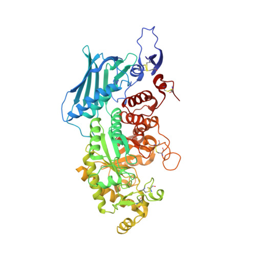
    β-N-acetyl-D-hexosaminidase has been postulated to have a specialized function. However, the structural basis of this specialization is not yet established. OfHex1, the enzyme from the Asian corn borer Ostrinia furnacalis (one of the most destructive pests) has previously been reported to function merely in chitin degradation. Here the vital role of OfHex1 during the pupation of O. furnacalis was revealed by RNA interference, and the crystal structures of OfHex1 and OfHex1 complexed with TMG-chitotriomycin were determined at 2.1 Å. The mechanism of selective inhibition by TMG-chitotriomycin was related to the existence of the +1 subsite at the active pocket of OfHex1 and a key residue, Trp(490), at this site. Mutation of Trp(490) to Ala led to a 2,277-fold decrease in sensitivity toward TMG-chitotriomycin as well as an 18-fold decrease in binding affinity for the substrate (GlcNAc)(2). Although the overall topology of the catalytic domain of OfHex1 shows a high similarity with the human and bacterial enzymes, OfHex1 is distinguished from these enzymes by large conformational changes linked to an "open-close" mechanism at the entrance of the active site, which is characterized by the "lid" residue, Trp(448). Mutation of Trp(448) to Ala or Phe resulted in a more than 1,000-fold loss in enzyme activity, due mainly to the effect on k(cat). The current work has increased our understanding of the structure-function relationship of OfHex1, shedding light on the structural basis that accounts for the specialized function of β-N-acetyl-D-hexosaminidase as well as making the development of species-specific pesticides a likely reality.


  • Organizational Affiliation
    • Department of Bioscience and Biotechnology, Dalian University of Technology, Dalian 116024, China.

Macromolecule Content 

  • Total Structure Weight: 66.38 kDa 
  • Atom Count: 5,097 
  • Modeled Residue Count: 572 
  • Deposited Residue Count: 572 
  • Unique protein chains: 1

Macromolecules

Find similar proteins by:|  3D Structure
Entity ID: 1
MoleculeChains  Sequence LengthOrganismDetailsImage
N-acetylglucosaminidase572Ostrinia furnacalisMutation(s): 0 
EC: 3.2.1.52
UniProt
Find proteins for Q06GJ0 (Ostrinia furnacalis)
Explore Q06GJ0 
Go to UniProtKB:  Q06GJ0
Entity Groups
Sequence Clusters30% Identity50% Identity70% Identity90% Identity95% Identity100% Identity
UniProt GroupQ06GJ0
Sequence Annotations
Expand
Reference Sequence

Oligosaccharides

Help  
Entity ID: 2
MoleculeChains Length2D Diagram GlycosylationD Interactions
2-deoxy-2-(trimethylammonio)-beta-D-glucopyranose-(1-4)-2-acetamido-2-deoxy-beta-D-glucopyranose-(1-4)-2-acetamido-2-deoxy-beta-D-glucopyranose-(1-4)-2-acetamido-2-deoxy-beta-D-glucopyranose
B
4N/AN/A
Glycosylation Resources
GlyTouCan: G26608LT
GlyCosmos: G26608LT

Biologically Interesting Molecules (External Reference) 

1 Unique

Experimental Data & Validation

Experimental Data

  • Method: X-RAY DIFFRACTION
  • Resolution: 2.10 Å
  • R-Value Work:  0.202 (Depositor), 0.217 (DCC) 
  • R-Value Observed: 0.202 (Depositor) 
Space Group: P 32 2 1
Unit Cell:
Length ( Å )Angle ( ˚ )
a = 108.5α = 90
b = 108.5β = 90
c = 175.57γ = 120
Software Package:
Software NamePurpose
HKL-2000data collection
CNSrefinement
REFMACrefinement
HKL-2000data reduction
HKL-2000data scaling
CNSphasing

Structure Validation

View Full Validation Report


Entry History 

Deposition Data

Revision History  (Full details and data files)

  • Version 1.0: 2010-11-24
    Type: Initial release
  • Version 1.1: 2011-07-13
    Changes: Version format compliance
  • Version 2.0: 2020-07-29
    Type: Remediation
    Reason: Carbohydrate remediation
    Changes: Atomic model, Data collection, Derived calculations, Non-polymer description, Structure summary
  • Version 2.1: 2023-11-01
    Changes: Data collection, Database references, Derived calculations, Refinement description, Structure summary
  • Version 2.2: 2024-10-09
    Changes: Structure summary