3NJQ

Crystal structure of Kaposi's sarcoma-associated herpesvirus protease in complex with dimer disruptor


Experimental Data Snapshot

  • Method: X-RAY DIFFRACTION
  • Resolution: 2.00 Å
  • R-Value Free: 0.248 
  • R-Value Work: 0.203 
  • R-Value Observed: 0.205 

wwPDB Validation   3D Report Full Report


Ligand Structure Quality Assessment 


This is version 1.2 of the entry. See complete history


Literature

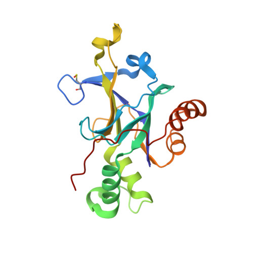
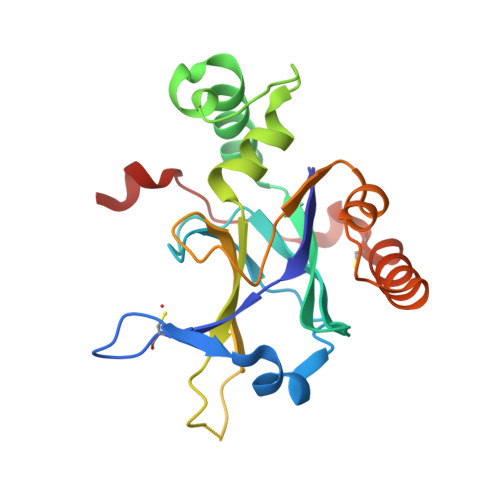
Enzyme inhibition by allosteric capture of an inactive conformation.

Lee, G.M.Shahian, T.Baharuddin, A.Gable, J.E.Craik, C.S.

(2011) J Mol Biol 411: 999-1016

  • DOI: https://doi.org/10.1016/j.jmb.2011.06.032
  • Primary Citation of Related Structures:  
    3NJQ

  • PubMed Abstract: 

    All members of the human herpesvirus protease (HHV Pr) family are active as weakly associating dimers but inactive as monomers. A small-molecule allosteric inhibitor of Kaposi's sarcoma-associated herpesvirus protease (KSHV Pr) traps the enzyme in an inactive monomeric state where the C-terminal helices are unfolded and the hydrophobic dimer interface is exposed. NMR titration studies demonstrate that the inhibitor binds to KSHV Pr monomers with low micromolar affinity. A 2.0-Å-resolution X-ray crystal structure of a C-terminal truncated KSHV Pr-inhibitor complex locates the binding pocket at the dimer interface and displays significant conformational perturbations at the active site, 15 Å from the allosteric site. NMR and CD data suggest that the small molecule inhibits human cytomegalovirus protease via a similar mechanism. As all HHV Prs are functionally and structurally homologous, the inhibitor represents a class of compounds that may be developed into broad-spectrum therapeutics that allosterically regulate enzymatic activity by disrupting protein-protein interactions.


  • Organizational Affiliation

    Department of Pharmaceutical Chemistry, University of California, San Francisco, CA 94158-2280, USA.


Macromolecules
Find similar proteins by:  (by identity cutoff)  |  3D Structure
Entity ID: 1
MoleculeChains Sequence LengthOrganismDetailsImage
ORF 17193Human herpesvirus 8 type MMutation(s): 0 
Gene Names: Rhadinovirus
UniProt
Find proteins for P88911 (Human herpesvirus 8 type M)
Explore P88911 
Go to UniProtKB:  P88911
Entity Groups  
Sequence Clusters30% Identity50% Identity70% Identity90% Identity95% Identity100% Identity
UniProt GroupP88911
Sequence Annotations
Expand
  • Reference Sequence
Find similar proteins by:  (by identity cutoff)  |  3D Structure
Entity ID: 2
MoleculeChains Sequence LengthOrganismDetailsImage
ORF 17193Human herpesvirus 8 type MMutation(s): 0 
Gene Names: Rhadinovirus
UniProt
Find proteins for P88911 (Human herpesvirus 8 type M)
Explore P88911 
Go to UniProtKB:  P88911
Entity Groups  
Sequence Clusters30% Identity50% Identity70% Identity90% Identity95% Identity100% Identity
UniProt GroupP88911
Sequence Annotations
Expand
  • Reference Sequence
Small Molecules
Ligands 4 Unique
IDChains Name / Formula / InChI Key2D Diagram3D Interactions
NJQ
Query on NJQ

Download Ideal Coordinates CCD File 
D [auth A],
I [auth B],
J [auth B]
3-benzyl-4-({[6-(cyclohexylmethyl)pyridin-2-yl]carbonyl}amino)benzoic acid
C27 H28 N2 O3
QBZUPKMHCWADDF-UHFFFAOYSA-N
SO4
Query on SO4

Download Ideal Coordinates CCD File 
E [auth B],
F [auth B],
G [auth B]
SULFATE ION
O4 S
QAOWNCQODCNURD-UHFFFAOYSA-L
GOL
Query on GOL

Download Ideal Coordinates CCD File 
H [auth B]GLYCEROL
C3 H8 O3
PEDCQBHIVMGVHV-UHFFFAOYSA-N
ACT
Query on ACT

Download Ideal Coordinates CCD File 
C [auth A]ACETATE ION
C2 H3 O2
QTBSBXVTEAMEQO-UHFFFAOYSA-M
Modified Residues  1 Unique
IDChains TypeFormula2D DiagramParent
CSO
Query on CSO
A
L-PEPTIDE LINKINGC3 H7 N O3 SCYS
Binding Affinity Annotations 
IDSourceBinding Affinity
NJQ Binding MOAD:  3NJQ Kd: 1700 (nM) from 1 assay(s)
PDBBind:  3NJQ Kd: 1.30e+4 (nM) from 1 assay(s)
Experimental Data & Validation

Experimental Data

  • Method: X-RAY DIFFRACTION
  • Resolution: 2.00 Å
  • R-Value Free: 0.248 
  • R-Value Work: 0.203 
  • R-Value Observed: 0.205 
  • Space Group: I 2 2 2
Unit Cell:
Length ( Å )Angle ( ˚ )
a = 69.317α = 90
b = 95.994β = 90
c = 119.244γ = 90
Software Package:
Software NamePurpose
DENZOdata reduction
SCALEPACKdata scaling
MOLREPphasing
REFMACrefinement
PDB_EXTRACTdata extraction
HKL-2000data collection
HKL-2000data reduction

Structure Validation

View Full Validation Report


Ligand Structure Quality Assessment 


Entry History 

Deposition Data

Revision History  (Full details and data files)

  • Version 1.0: 2011-08-17
    Type: Initial release
  • Version 1.1: 2011-08-31
    Changes: Database references
  • Version 1.2: 2017-11-08
    Changes: Refinement description