


Crystal structure of carbonic anhydrase II in complex with a Nir inhibitor
Temperini, C., Cecchi, A.To be published.
Experimental Data Snapshot
Starting Model: experimental
View more details
Entity ID: 1 | |||||
|---|---|---|---|---|---|
| Molecule | Chains | Sequence Length | Organism | Details | Image |
| Carbonic anhydrase 2 | 260 | Homo sapiens | Mutation(s): 0 EC: 4.2.1.1 (PDB Primary Data), 4.2.1.69 (UniProt) | 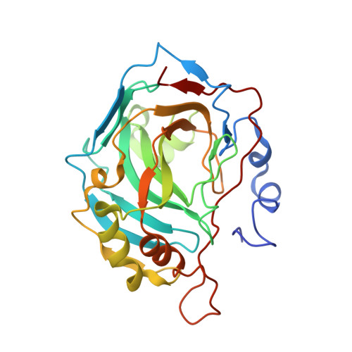 | |
UniProt & NIH Common Fund Data Resources | |||||
PHAROS: P00918 GTEx: ENSG00000104267 | |||||
Entity Groups | |||||
| Sequence Clusters | 30% Identity50% Identity70% Identity90% Identity95% Identity100% Identity | ||||
| UniProt Group | P00918 | ||||
Sequence AnnotationsExpand | |||||
Reference Sequence | |||||
| Ligands 3 Unique | |||||
|---|---|---|---|---|---|
| ID | Chains | Name / Formula / InChI Key | 2D Diagram | 3D Interactions | |
| TE2 Download:Ideal Coordinates CCD File | D [auth A] | 3-(4-sulfamoylphenyl)-N-[6-({(6Z)-2-[(2Z)-2-(1,3,3-trimethyl-1,3-dihydro-2H-indol-2-ylidene)ethyl]-6-[(2E)-2-(1,3,3-trimethyl-1,3-dihydro-2H-indol-2-ylidene)ethylidene]cyclohex-1-en-1-yl}amino)hexyl]propanamide C47 H61 N5 O3 S KNDSETYOKYYUHZ-OLEWYFQESA-N |  | ||
| HG Download:Ideal Coordinates CCD File | C [auth A] | MERCURY (II) ION Hg BQPIGGFYSBELGY-UHFFFAOYSA-N |  | ||
| ZN Download:Ideal Coordinates CCD File | B [auth A] | ZINC ION Zn PTFCDOFLOPIGGS-UHFFFAOYSA-N |  | ||
| Length ( Å ) | Angle ( ˚ ) |
|---|---|
| a = 42.07 | α = 90 |
| b = 41.54 | β = 104.45 |
| c = 72.39 | γ = 90 |
| Software Name | Purpose |
|---|---|
| CrysalisPro | data collection |
| AMoRE | phasing |
| REFMAC | refinement |
| CrysalisPro | data reduction |
| SCALEPACK | data scaling |


