3NIF | pdb_00003nif

The Closed Headpiece of Integrin IIb 3 and its Complex with an IIb 3 -Specific Antagonist That Does Not Induce Opening


Experimental Data Snapshot

  • Method: X-RAY DIFFRACTION
  • Resolution: 2.40 Å
  • R-Value Free: 
    0.216 (Depositor), 0.224 (DCC) 
  • R-Value Work: 
    0.176 (Depositor) 
  • R-Value Observed: 
    0.176 (Depositor) 

Starting Model: experimental
View more details

wwPDB Validation 3D Report Full Report

Validation slider image for 3NIF

Ligand Structure Quality Assessment 


This is version 2.2 of the entry. See complete history

Literature

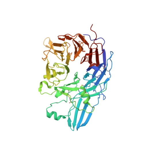
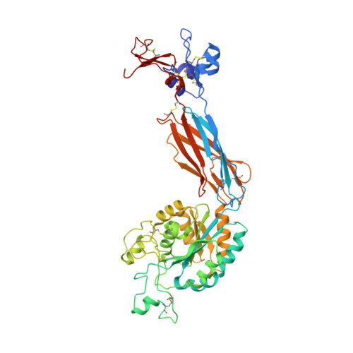
Closed headpiece of integrin {alpha}IIb{beta}3 and its complex with an {alpha}IIb{beta}3-specific antagonist that does not induce opening.

Zhu, J.Zhu, J.Negri, A.Provasi, D.Filizola, M.Coller, B.S.Springer, T.A.

(2010) Blood 116: 5050-5059

  • DOI: https://doi.org/10.1182/blood-2010-04-281154
  • Primary Citation Related Structures: 
    3NID, 3NIF, 3NIG

  • PubMed Abstract: 

    The platelet integrin α(IIb)β(3) is essential for hemostasis and thrombosis through its binding of adhesive plasma proteins. We have determined crystal structures of the α(IIb)β(3) headpiece in the absence of ligand and after soaking in RUC-1, a novel small molecule antagonist. In the absence of ligand, the α(IIb)β(3) headpiece is in a closed conformation, distinct from the open conformation visualized in presence of Arg-Gly-Asp (RGD) antagonists. In contrast to RGD antagonists, RUC-1 binds only to the α(IIb) subunit. Molecular dynamics revealed nearly identical binding. Two species-specific residues, α(IIb) Y190 and α(IIb) D232, in the RUC-1 binding site were confirmed as important by mutagenesis. In sharp contrast to RGD-based antagonists, RUC-1 did not induce α(IIb)β(3) to adopt an open conformation, as determined by gel filtration and dynamic light scattering. These studies provide insights into the factors that regulate integrin headpiece opening, and demonstrate the molecular basis for a novel mechanism of integrin antagonism.


  • Organizational Affiliation
    • Immune Disease Institute, Children's Hospital Boston, and Department of Pathology, Harvard Medical School, Boston, MA 02115, USA.

Macromolecule Content 

  • Total Structure Weight: 303.3 kDa 
  • Atom Count: 21,915 
  • Modeled Residue Count: 2,703 
  • Deposited Residue Count: 2,726 
  • Unique protein chains: 4

Macromolecules

Find similar proteins by:|  3D Structure
Entity ID: 1
MoleculeChains  Sequence LengthOrganismDetailsImage
Integrin alphaIIB beta3
A, C
457Homo sapiensMutation(s): 0 
Gene Names: ITGA2BGP2BITGAB
UniProt & NIH Common Fund Data Resources
Find proteins for P08514 (Homo sapiens)
Explore P08514 
Go to UniProtKB:  P08514
PHAROS:  P08514
GTEx:  ENSG00000005961 
Entity Groups
Sequence Clusters30% Identity50% Identity70% Identity90% Identity95% Identity100% Identity
UniProt GroupP08514
Sequence Annotations
Expand
Reference Sequence
Find similar proteins by:|  3D Structure
Entity ID: 2
MoleculeChains  Sequence LengthOrganismDetailsImage
Integrin beta-3
B, D
471Homo sapiensMutation(s): 0 
Gene Names: ITGB3GP3A
UniProt & NIH Common Fund Data Resources
Find proteins for P05106 (Homo sapiens)
Explore P05106 
Go to UniProtKB:  P05106
PHAROS:  P05106
GTEx:  ENSG00000259207 
Entity Groups
Sequence Clusters30% Identity50% Identity70% Identity90% Identity95% Identity100% Identity
UniProt GroupP05106
Glycosylation
Glycosylation Sites: 3Go to GlyGen: P05106-1
Sequence Annotations
Expand
Reference Sequence
Find similar proteins by:|  3D Structure
Entity ID: 3
MoleculeChains  Sequence LengthOrganismDetailsImage
Monoclonal antibody 10E5 heavy chainE,
G [auth H]
221Mus musculusMutation(s): 0 
Find similar proteins by:|  3D Structure
Entity ID: 4
MoleculeChains  Sequence LengthOrganismDetailsImage
Monoclonal antibody 10E5 light chainF,
H [auth L]
214Mus musculusMutation(s): 0 

Oligosaccharides

Help  
Entity ID: 5
MoleculeChains Length2D Diagram GlycosylationD Interactions
alpha-D-mannopyranose-(1-3)-alpha-D-mannopyranose-(1-4)-2-acetamido-2-deoxy-beta-D-glucopyranose-(1-4)-2-acetamido-2-deoxy-beta-D-glucopyranoseI [auth G]4N-Glycosylation
Glycosylation Resources
GlyTouCan: G48068RF
GlyCosmos: G48068RF
GlyGen: G48068RF
Entity ID: 6
MoleculeChains Length2D Diagram GlycosylationD Interactions
2-acetamido-2-deoxy-beta-D-glucopyranose-(1-4)-2-acetamido-2-deoxy-beta-D-glucopyranoseJ [auth I],
L [auth K]
2N-Glycosylation
Glycosylation Resources
GlyTouCan: G42666HT
GlyCosmos: G42666HT
GlyGen: G42666HT
Entity ID: 7
MoleculeChains Length2D Diagram GlycosylationD Interactions
alpha-D-mannopyranose-(1-4)-2-acetamido-2-deoxy-beta-D-glucopyranose-(1-4)-2-acetamido-2-deoxy-beta-D-glucopyranoseK [auth J]3N-Glycosylation
Glycosylation Resources
GlyTouCan: G62182OO
GlyCosmos: G62182OO
GlyGen: G62182OO

Small Molecules

Ligands 6 Unique
IDChains Name / Formula / InChI Key2D Diagram3D Interactions
NIF

Query on NIF


Download:Ideal Coordinates CCD File
MA [auth C],
Q [auth A]
2-ethyl-7-piperazin-1-yl-5H-[1,3,4]thiadiazolo[3,2-a]pyrimidin-5-one
C11 H15 N5 O S
WUYNDJNASMITGA-UHFFFAOYSA-N
NAG

Query on NAG


Download:Ideal Coordinates CCD File
FA [auth B],
WA [auth D]
2-acetamido-2-deoxy-beta-D-glucopyranose
C8 H15 N O6
OVRNDRQMDRJTHS-FMDGEEDCSA-N
SO4

Query on SO4


Download:Ideal Coordinates CCD File
AA [auth A]
AB [auth D]
BA [auth A]
BB [auth H]
CB [auth L]
AA [auth A],
AB [auth D],
BA [auth A],
BB [auth H],
CB [auth L],
DB [auth L],
HA [auth B],
NA [auth C],
OA [auth C],
PA [auth C],
QA [auth C],
RA [auth C],
SA [auth C],
X [auth A],
Y [auth A],
YA [auth D],
Z [auth A],
ZA [auth D]
SULFATE ION
O4 S
QAOWNCQODCNURD-UHFFFAOYSA-L
GOL

Query on GOL


Download:Ideal Coordinates CCD File
GA [auth B]
R [auth A]
S [auth A]
T [auth A]
U [auth A]
GA [auth B],
R [auth A],
S [auth A],
T [auth A],
U [auth A],
V [auth A],
W [auth A],
XA [auth D]
GLYCEROL
C3 H8 O3
PEDCQBHIVMGVHV-UHFFFAOYSA-N
CA

Query on CA


Download:Ideal Coordinates CCD File
DA [auth B]
EA [auth B]
IA [auth C]
JA [auth C]
KA [auth C]
DA [auth B],
EA [auth B],
IA [auth C],
JA [auth C],
KA [auth C],
LA [auth C],
M [auth A],
N [auth A],
O [auth A],
P [auth A],
UA [auth D],
VA [auth D]
CALCIUM ION
Ca
BHPQYMZQTOCNFJ-UHFFFAOYSA-N
MG

Query on MG


Download:Ideal Coordinates CCD File
CA [auth B],
TA [auth D]
MAGNESIUM ION
Mg
JLVVSXFLKOJNIY-UHFFFAOYSA-N
Binding Affinity Annotations 
IDSourceBinding Affinity
NIF BindingDB:  3NIF IC50: min: 9600, max: 1.30e+4 (nM) from 3 assay(s)

Experimental Data & Validation

Experimental Data

  • Method: X-RAY DIFFRACTION
  • Resolution: 2.40 Å
  • R-Value Free:  0.216 (Depositor), 0.224 (DCC) 
  • R-Value Work:  0.176 (Depositor) 
  • R-Value Observed: 0.176 (Depositor) 
Space Group: P 21 21 2
Unit Cell:
Length ( Å )Angle ( ˚ )
a = 259.53α = 90
b = 144.28β = 90
c = 104.35γ = 90
Software Package:
Software NamePurpose
REFMACrefinement

Structure Validation

View Full Validation Report


Ligand Structure Quality Assessment 


Entry History 

Deposition Data

Revision History  (Full details and data files)

  • Version 1.0: 2010-12-22
    Type: Initial release
  • Version 1.1: 2011-07-13
    Changes: Version format compliance
  • Version 2.0: 2020-07-29
    Type: Remediation
    Reason: Carbohydrate remediation
    Changes: Advisory, Atomic model, Data collection, Derived calculations, Structure summary
  • Version 2.1: 2023-09-06
    Changes: Data collection, Database references, Refinement description, Structure summary
  • Version 2.2: 2024-10-16
    Changes: Structure summary