3NCT

X-ray crystal structure of the bacterial conjugation factor PsiB, a negative regulator of reca


Experimental Data Snapshot

  • Method: X-RAY DIFFRACTION
  • Resolution: 2.20 Å
  • R-Value Free: 0.210 
  • R-Value Work: 0.185 
  • R-Value Observed: 0.186 

wwPDB Validation   3D Report Full Report


This is version 1.1 of the entry. See complete history


Literature

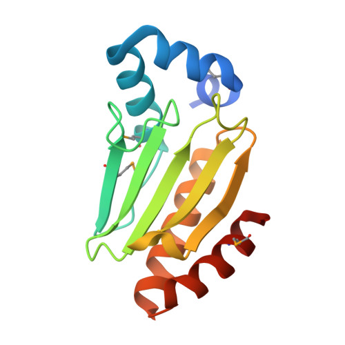
X-ray crystal structure of the bacterial conjugation factor PsiB, a negative regulator of RecA.

Petrova, V.Satyshur, K.A.George, N.P.McCaslin, D.Cox, M.M.Keck, J.L.

(2010) J Biol Chem 285: 30615-30621

  • DOI: https://doi.org/10.1074/jbc.M110.152298
  • Primary Citation of Related Structures:  
    3NCT

  • PubMed Abstract: 

    During bacterial conjugation, genetic material from one cell is transferred to another as single-stranded DNA. The introduction of single-stranded DNA into the recipient cell would ordinarily trigger a potentially deleterious transcriptional response called SOS, which is initiated by RecA protein filaments formed on the DNA. During F plasmid conjugation, however, the SOS response is suppressed by PsiB, an F-plasmid-encoded protein that binds and sequesters free RecA to prevent filament formation. Among the many characterized RecA modulator proteins, PsiB is unique in using sequestration as an inhibitory mechanism. We describe the crystal structure of PsiB from the Escherichia coli F plasmid. The stucture of PsiB is surprisingly similar to CapZ, a eukaryotic actin filament capping protein. Structure-directed neutralization of electronegative surfaces on PsiB abrogates RecA inhibition whereas neutralization of an electropositive surface element enhances PsiB inhibition of RecA. Together, these studies provide a first molecular view of PsiB and highlight its use as a reagent in studies of RecA activity.


  • Organizational Affiliation

    University of Wisconsin, Madison, Wisconsin 53706-1532, USA.


Macromolecules
Find similar proteins by:  (by identity cutoff)  |  3D Structure
Entity ID: 1
MoleculeChains Sequence LengthOrganismDetailsImage
Protein psiB
A, B, C, D
144Escherichia coliMutation(s): 0 
Gene Names: ECOK12F063psiB
UniProt
Find proteins for P10031 (Escherichia coli (strain K12))
Explore P10031 
Go to UniProtKB:  P10031
Entity Groups  
Sequence Clusters30% Identity50% Identity70% Identity90% Identity95% Identity100% Identity
UniProt GroupP10031
Sequence Annotations
Expand
  • Reference Sequence
Small Molecules
Modified Residues  1 Unique
IDChains TypeFormula2D DiagramParent
MSE
Query on MSE
A, B, C, D
L-PEPTIDE LINKINGC5 H11 N O2 SeMET
Experimental Data & Validation

Experimental Data

  • Method: X-RAY DIFFRACTION
  • Resolution: 2.20 Å
  • R-Value Free: 0.210 
  • R-Value Work: 0.185 
  • R-Value Observed: 0.186 
  • Space Group: P 43 21 2
Unit Cell:
Length ( Å )Angle ( ˚ )
a = 93.815α = 90
b = 93.815β = 90
c = 164.827γ = 90
Software Package:
Software NamePurpose
ADSCdata collection
SOLVEphasing
REFMACrefinement
HKL-2000data reduction
HKL-2000data scaling

Structure Validation

View Full Validation Report


Entry History 

Deposition Data

Revision History  (Full details and data files)

  • Version 1.0: 2010-07-21
    Type: Initial release
  • Version 1.1: 2011-07-13
    Changes: Version format compliance