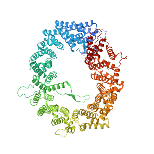
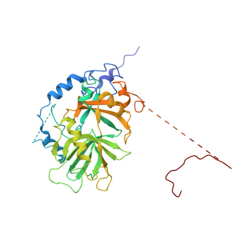
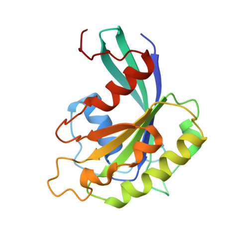
3NC0

Crystal structure of the HIV-1 Rev NES-CRM1-RanGTP nuclear export complex (crystal II)


Experimental Data Snapshot

  • Method: X-RAY DIFFRACTION
  • Resolution: 2.90 Å
  • R-Value Free: 0.295 
  • R-Value Work: 0.242 
  • R-Value Observed: 0.245 

Starting Model: experimental
View more details

wwPDB Validation   3D Report Full Report


Ligand Structure Quality Assessment 


This is version 1.3 of the entry. See complete history


Literature

NES consensus redefined by structures of PKI-type and Rev-type nuclear export signals bound to CRM1.

Guttler, T.Madl, T.Neumann, P.Deichsel, D.Corsini, L.Monecke, T.Ficner, R.Sattler, M.Gorlich, D.

(2010) Nat Struct Mol Biol 17: 1367-1376

  • DOI: https://doi.org/10.1038/nsmb.1931
  • Primary Citation of Related Structures:  
    2L1L, 3NBY, 3NBZ, 3NC0, 3NC1

  • PubMed Abstract: 

    Classic nuclear export signals (NESs) confer CRM1-dependent nuclear export. Here we present crystal structures of the RanGTP-CRM1 complex alone and bound to the prototypic PKI or HIV-1 Rev NESs. These NESs differ markedly in the spacing of their key hydrophobic (Φ) residues, yet CRM1 recognizes them with the same rigid set of five Φ pockets. The different Φ spacings are compensated for by different conformations of the bound NESs: in the case of PKI, an α-helical conformation, and in the case of Rev, an extended conformation with a critical proline docking into a Φ pocket. NMR analyses of CRM1-bound and CRM1-free PKI NES suggest that CRM1 selects NES conformers that pre-exist in solution. Our data lead to a new structure-based NES consensus, and explain why NESs differ in their affinities for CRM1 and why supraphysiological NESs bind the exportin so tightly.


  • Organizational Affiliation

    Max-Planck-Institut für Biophysikalische Chemie, Göttingen, Germany.


Macromolecules
Find similar proteins by:  (by identity cutoff)  |  3D Structure
Entity ID: 1
MoleculeChains Sequence LengthOrganismDetailsImage
Exportin-1
A, D
1,073Mus musculusMutation(s): 0 
Gene Names: Crm1Xpo1Xpo1 (GeneID: 103573)
UniProt & NIH Common Fund Data Resources
Find proteins for Q6P5F9 (Mus musculus)
Explore Q6P5F9 
Go to UniProtKB:  Q6P5F9
IMPC:  MGI:2144013
Entity Groups  
Sequence Clusters30% Identity50% Identity70% Identity90% Identity95% Identity100% Identity
UniProt GroupQ6P5F9
Sequence Annotations
Expand
  • Reference Sequence
Find similar proteins by:  (by identity cutoff)  |  3D Structure
Entity ID: 2
MoleculeChains Sequence LengthOrganismDetailsImage
Snurportin-1
B, E
362Homo sapiensMutation(s): 0 
Gene Names: SNUPN (GeneID: 10073)RNUT1SNUPNSPN1
UniProt & NIH Common Fund Data Resources
Find proteins for O95149 (Homo sapiens)
Explore O95149 
Go to UniProtKB:  O95149
GTEx:  ENSG00000169371 
Entity Groups  
Sequence Clusters30% Identity50% Identity70% Identity90% Identity95% Identity100% Identity
UniProt GroupO95149
Sequence Annotations
Expand
  • Reference Sequence
Find similar proteins by:  (by identity cutoff)  |  3D Structure
Entity ID: 3
MoleculeChains Sequence LengthOrganismDetailsImage
GTP-binding nuclear protein Ran
C, F
176Homo sapiensMutation(s): 1 
Gene Names: ARA24OK/SW-cl.81RANRAN (GeneID: 5901)
UniProt & NIH Common Fund Data Resources
Find proteins for P62826 (Homo sapiens)
Explore P62826 
Go to UniProtKB:  P62826
PHAROS:  P62826
GTEx:  ENSG00000132341 
Entity Groups  
Sequence Clusters30% Identity50% Identity70% Identity90% Identity95% Identity100% Identity
UniProt GroupP62826
Sequence Annotations
Expand
  • Reference Sequence
Small Molecules
Ligands 5 Unique
IDChains Name / Formula / InChI Key2D Diagram3D Interactions
GTP
Query on GTP

Download Ideal Coordinates CCD File 
EA [auth F],
Q [auth C]
GUANOSINE-5'-TRIPHOSPHATE
C10 H16 N5 O14 P3
XKMLYUALXHKNFT-UUOKFMHZSA-N
PEG
Query on PEG

Download Ideal Coordinates CCD File 
AA [auth D]
GA [auth L]
J [auth A]
K [auth A]
L [auth A]
AA [auth D],
GA [auth L],
J [auth A],
K [auth A],
L [auth A],
M [auth A],
T [auth C],
V [auth D],
W [auth D],
X [auth D],
Y [auth D],
Z [auth D]
DI(HYDROXYETHYL)ETHER
C4 H10 O3
MTHSVFCYNBDYFN-UHFFFAOYSA-N
IPH
Query on IPH

Download Ideal Coordinates CCD File 
O [auth B]PHENOL
C6 H6 O
ISWSIDIOOBJBQZ-UHFFFAOYSA-N
GOL
Query on GOL

Download Ideal Coordinates CCD File 
BA [auth D]
CA [auth E]
DA [auth E]
G [auth A]
H [auth A]
BA [auth D],
CA [auth E],
DA [auth E],
G [auth A],
H [auth A],
I [auth A],
N [auth A],
P [auth B],
S [auth C],
U [auth D]
GLYCEROL
C3 H8 O3
PEDCQBHIVMGVHV-UHFFFAOYSA-N
MG
Query on MG

Download Ideal Coordinates CCD File 
FA [auth F],
R [auth C]
MAGNESIUM ION
Mg
JLVVSXFLKOJNIY-UHFFFAOYSA-N
Experimental Data & Validation

Experimental Data

  • Method: X-RAY DIFFRACTION
  • Resolution: 2.90 Å
  • R-Value Free: 0.295 
  • R-Value Work: 0.242 
  • R-Value Observed: 0.245 
  • Space Group: P 1 21 1
Unit Cell:
Length ( Å )Angle ( ˚ )
a = 73.262α = 90
b = 225.895β = 100.75
c = 163.984γ = 90
Software Package:
Software NamePurpose
XSCALEdata processing
PHENIXrefinement
PDB_EXTRACTdata extraction
MAR345dtbdata collection
XDSdata reduction
XSCALEdata scaling
CNSphasing

Structure Validation

View Full Validation Report


Ligand Structure Quality Assessment 


Entry History 

Deposition Data

Revision History  (Full details and data files)

  • Version 1.0: 2010-10-27
    Type: Initial release
  • Version 1.1: 2011-07-13
    Changes: Version format compliance
  • Version 1.2: 2021-10-06
    Changes: Database references, Derived calculations
  • Version 1.3: 2023-09-06
    Changes: Data collection, Refinement description