3N9K

F229A/E292S Double Mutant of Exo-beta-1,3-glucanase from Candida albicans in Complex with Laminaritriose at 1.7 A


Experimental Data Snapshot

  • Method: X-RAY DIFFRACTION
  • Resolution: 1.70 Å
  • R-Value Free: 0.197 
  • R-Value Work: 0.152 
  • R-Value Observed: 0.154 

wwPDB Validation   3D Report Full Report


This is version 2.1 of the entry. See complete history


Literature

Carbohydrate binding sites in Candida albicans exo-beta-1,3-glucanase and the role of the Phe-Phe 'clamp' at the active site entrance

Patrick, W.M.Nakatani, Y.Cutfield, S.M.Sharpe, M.L.Ramsay, R.J.Cutfield, J.F.

(2010) FEBS J 277: 4549-4561

  • DOI: https://doi.org/10.1111/j.1742-4658.2010.07869.x
  • Primary Citation of Related Structures:  
    2PC8, 2PF0, 3N9K, 3O6A

  • PubMed Abstract: 

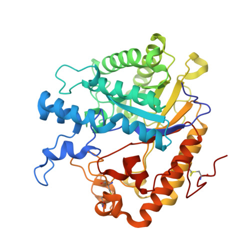
    Candida albicans exo-β-1,3-glucanase (Exg; EC 3.2.1.58) is implicated in cell wall β-D-glucan remodelling through its glucosyl hydrolase and/or transglucosylase activities. A pair of antiparallel phenylalanyl residues (F144 and F258) flank the entrance to the active site pocket. Various Exg mutants were studied using steady-state kinetics and crystallography aiming to understand the roles played by these residues in positioning the β-1,3-D-glucan substrate. Mutations at the Phe-Phe entranceway demonstrated the requirement for double-sided CH/π interactions at the +1 subsite, and the necessity for phenylalanine rather than tyrosine or tryptophan. The Tyr-Tyr double mutations introduced ordered water molecules into the entranceway. A third Phe residue (F229) nearby was evaluated as a possible +2 subsite. The inactive double mutant E292S/F229A complexed with laminaritriose has provided the first picture of substrate binding to Exg and demonstrated how the Phe-Phe arrangement acts as a clamp at the +1 subsite. The terminal sugar at the -1 site showed displacement from the position of a monosaccharide analogue with interchange of water molecules and sugar hydroxyls. An unexpected additional glucose binding site, well removed from the active site, was revealed. This site may enable Exg to associate with the branched glucan structure of the C. albicans cell wall.


  • Organizational Affiliation

    Department of Biochemistry, University of Otago, Dunedin, New Zealand.


Macromolecules
Find similar proteins by:  (by identity cutoff)  |  3D Structure
Entity ID: 1
MoleculeChains Sequence LengthOrganismDetailsImage
Glucan 1,3-beta-glucosidase399Candida albicansMutation(s): 2 
Gene Names: XOG
EC: 3.2.1.58
UniProt
Find proteins for P29717 (Candida albicans (strain SC5314 / ATCC MYA-2876))
Explore P29717 
Go to UniProtKB:  P29717
Entity Groups  
Sequence Clusters30% Identity50% Identity70% Identity90% Identity95% Identity100% Identity
UniProt GroupP29717
Sequence Annotations
Expand
  • Reference Sequence
Oligosaccharides

Help

Entity ID: 2
MoleculeChains Length2D Diagram Glycosylation3D Interactions
beta-D-glucopyranose-(1-3)-beta-D-glucopyranose-(1-3)-beta-D-glucopyranose
B
3N/A
Glycosylation Resources
GlyTouCan:  G00024MO
GlyCosmos:  G00024MO
GlyGen:  G00024MO
Entity ID: 3
MoleculeChains Length2D Diagram Glycosylation3D Interactions
beta-D-glucopyranose-(1-3)-beta-D-glucopyranose
C
2N/A
Glycosylation Resources
GlyTouCan:  G36535HU
GlyCosmos:  G36535HU
Small Molecules
Ligands 1 Unique
IDChains Name / Formula / InChI Key2D Diagram3D Interactions
CA
Query on CA

Download Ideal Coordinates CCD File 
D [auth A]CALCIUM ION
Ca
BHPQYMZQTOCNFJ-UHFFFAOYSA-N
Biologically Interesting Molecules (External Reference) 1 Unique
Experimental Data & Validation

Experimental Data

  • Method: X-RAY DIFFRACTION
  • Resolution: 1.70 Å
  • R-Value Free: 0.197 
  • R-Value Work: 0.152 
  • R-Value Observed: 0.154 
  • Space Group: P 21 21 21
Unit Cell:
Length ( Å )Angle ( ˚ )
a = 58.725α = 90
b = 64.397β = 90
c = 94.866γ = 90
Software Package:
Software NamePurpose
CrystalCleardata collection
MOLREPphasing
REFMACrefinement
XDSdata reduction
SCALAdata scaling

Structure Validation

View Full Validation Report


Entry History 

Deposition Data

Revision History  (Full details and data files)

  • Version 1.0: 2010-09-15
    Type: Initial release
  • Version 1.1: 2011-07-13
    Changes: Version format compliance
  • Version 2.0: 2020-07-29
    Type: Remediation
    Reason: Carbohydrate remediation
    Changes: Atomic model, Data collection, Database references, Derived calculations, Structure summary
  • Version 2.1: 2023-11-01
    Changes: Data collection, Database references, Refinement description, Structure summary