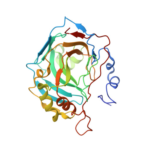
3N4B

Crystal structure of human carbonic anhydrase II in complex with a benzenesulfonamide inhibitor


Experimental Data Snapshot

  • Method: X-RAY DIFFRACTION
  • Resolution: 1.60 Å
  • R-Value Free: 0.170 
  • R-Value Work: 0.141 
  • R-Value Observed: 0.143 

Starting Model: experimental
View more details

wwPDB Validation   3D Report Full Report


Ligand Structure Quality Assessment 


This is version 1.3 of the entry. See complete history


Literature

Selective hydrophobic pocket binding observed within the carbonic anhydrase II active site accommodate different 4-substituted-ureido-benzenesulfonamides and correlate to inhibitor potency.

Pacchiano, F.Aggarwal, M.Avvaru, B.S.Robbins, A.H.Scozzafava, A.McKenna, R.Supuran, C.T.

(2010) Chem Commun (Camb) 46: 8371-8373

  • DOI: https://doi.org/10.1039/c0cc02707c
  • Primary Citation of Related Structures:  
    3MZC, 3N0N, 3N2P, 3N3J, 3N4B

  • PubMed Abstract: 

    4-Substituted-ureido benzenesulfonamides showing inhibitory activity against carbonic anhydrase (CA, EC 4.2.1.1) II between 3.3-226 nM were crystallized in complex with the enzyme. Hydrophobic interactions between the scaffold of the inhibitors in different hydrophobic pockets of the enzyme were observed, explaining the diverse inhibitory range of these derivatives.


  • Organizational Affiliation

    Department of Biochemistry and Molecular Biology, College of Medicine, University of Florida, Box 100245, Gainesville, Florida 32610, USA.


Macromolecules
Find similar proteins by:  (by identity cutoff)  |  3D Structure
Entity ID: 1
MoleculeChains Sequence LengthOrganismDetailsImage
Carbonic anhydrase 2260Homo sapiensMutation(s): 0 
Gene Names: HCA2
EC: 4.2.1.1
UniProt & NIH Common Fund Data Resources
Find proteins for P00918 (Homo sapiens)
Explore P00918 
Go to UniProtKB:  P00918
PHAROS:  P00918
GTEx:  ENSG00000104267 
Entity Groups  
Sequence Clusters30% Identity50% Identity70% Identity90% Identity95% Identity100% Identity
UniProt GroupP00918
Sequence Annotations
Expand
  • Reference Sequence
Small Molecules
Binding Affinity Annotations 
IDSourceBinding Affinity
WWZ BindingDB:  3N4B Ki: min: 96, max: 960 (nM) from 2 assay(s)
PDBBind:  3N4B Ki: 96 (nM) from 1 assay(s)
Experimental Data & Validation

Experimental Data

  • Method: X-RAY DIFFRACTION
  • Resolution: 1.60 Å
  • R-Value Free: 0.170 
  • R-Value Work: 0.141 
  • R-Value Observed: 0.143 
  • Space Group: P 1 21 1
Unit Cell:
Length ( Å )Angle ( ˚ )
a = 42.465α = 90
b = 41.414β = 104.13
c = 72.037γ = 90
Software Package:
Software NamePurpose
DENZOdata reduction
SCALEPACKdata scaling
PHENIXrefinement
PDB_EXTRACTdata extraction
CrystalCleardata collection
HKL-2000data reduction
HKL-2000data scaling
PHENIXphasing

Structure Validation

View Full Validation Report


Ligand Structure Quality Assessment 


Entry History 

Deposition Data

Revision History  (Full details and data files)

  • Version 1.0: 2011-03-09
    Type: Initial release
  • Version 1.1: 2011-07-13
    Changes: Version format compliance
  • Version 1.2: 2017-11-08
    Changes: Refinement description
  • Version 1.3: 2023-09-06
    Changes: Data collection, Database references, Derived calculations, Refinement description