3N41

Crystal structure of the mature envelope glycoprotein complex (spontaneous cleavage) of Chikungunya virus.


Experimental Data Snapshot

  • Method: X-RAY DIFFRACTION
  • Resolution: 3.01 Å
  • R-Value Free: 0.275 
  • R-Value Work: 0.252 
  • R-Value Observed: 0.253 

wwPDB Validation   3D Report Full Report


Ligand Structure Quality Assessment 


This is version 2.0 of the entry. See complete history


Literature

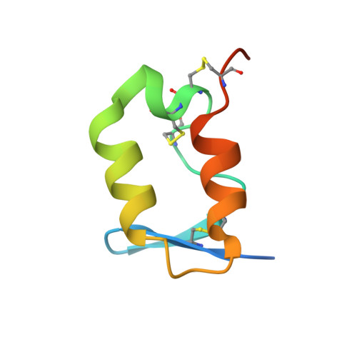
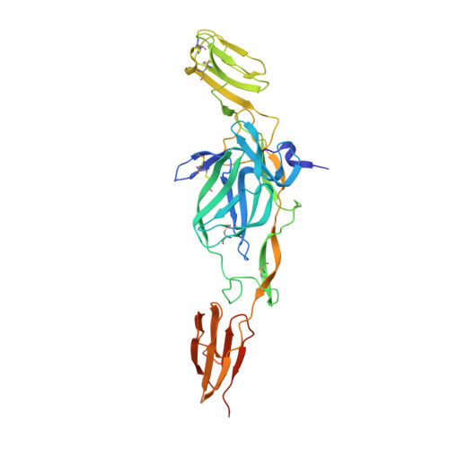
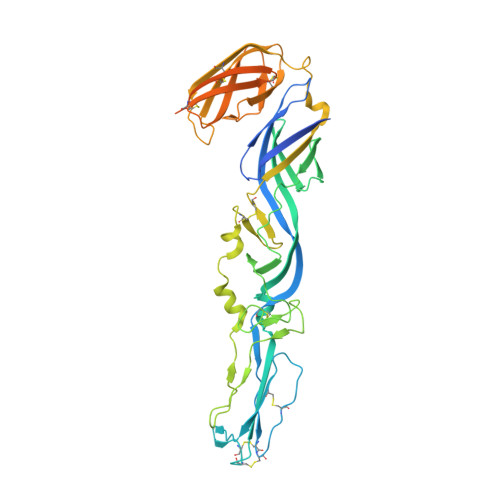
Glycoprotein organization of Chikungunya virus particles revealed by X-ray crystallography.

Voss, J.E.Vaney, M.C.Duquerroy, S.Vonrhein, C.Girard-Blanc, C.Crublet, E.Thompson, A.Bricogne, G.Rey, F.A.

(2010) Nature 468: 709-712

  • DOI: 10.1038/nature09555
  • Primary Citation of Related Structures:  
    2XFB, 2XFC, 3N40, 3N41, 3N42, 3N43, 3N44

  • PubMed Abstract: 
  • Chikungunya virus (CHIKV) is an emerging mosquito-borne alphavirus that has caused widespread outbreaks of debilitating human disease in the past five years. CHIKV invasion of susceptible cells is mediated by two viral glycoproteins, E1 and E2, which carry the main antigenic determinants and form an icosahedral shell at the virion surface ...

    Chikungunya virus (CHIKV) is an emerging mosquito-borne alphavirus that has caused widespread outbreaks of debilitating human disease in the past five years. CHIKV invasion of susceptible cells is mediated by two viral glycoproteins, E1 and E2, which carry the main antigenic determinants and form an icosahedral shell at the virion surface. Glycoprotein E2, derived from furin cleavage of the p62 precursor into E3 and E2, is responsible for receptor binding, and E1 for membrane fusion. In the context of a concerted multidisciplinary effort to understand the biology of CHIKV, here we report the crystal structures of the precursor p62-E1 heterodimer and of the mature E3-E2-E1 glycoprotein complexes. The resulting atomic models allow the synthesis of a wealth of genetic, biochemical, immunological and electron microscopy data accumulated over the years on alphaviruses in general. This combination yields a detailed picture of the functional architecture of the 25 MDa alphavirus surface glycoprotein shell. Together with the accompanying report on the structure of the Sindbis virus E2-E1 heterodimer at acidic pH (ref. 3), this work also provides new insight into the acid-triggered conformational change on the virus particle and its inbuilt inhibition mechanism in the immature complex.


    Related Citations: 
    • Structure and interactions at the viral surface of the envelope protein E1 of Semliki Forest virus.
      Roussel, A., Lescar, J., Vaney, M.C., Wengler, G., Rey, F.A.
      (2006) Structure 14: 75
    • Conformational change and protein-protein interactions of the fusion protein of Semliki Forest virus.
      Gibbons, D.L., Vaney, M.C., Roussel, A., Vigouroux, A., Reilly, B., Lepault, J., Kielian, M., Rey, F.A.
      (2004) Nature 427: 320
    • The Fusion glycoprotein shell of Semliki Forest virus: an icosahedral assembly primed for fusogenic activation at endosomal pH.
      Lescar, J., Roussel, A., Wien, M.W., Navaza, J., Fuller, S.D., Wengler, G., Rey, F.A.
      (2001) Cell 105: 137
    • Structural Changes of Envelope Proteins During Alphavirus Fusion.
      Li, L., Jose, J., Xiang, Y., Kuhn, R.J., Rossmann, M.G.
      () To be published --: --

    Organizational Affiliation

    Institut Pasteur, Département de Virologie, Unité de Virologie Structurale, 25 rue du Dr Roux, 75724 Paris Cedex 15, France.


Macromolecules
Find similar proteins by:  (by identity cutoff)  |  3D Structure
Entity ID: 1
MoleculeChainsSequence LengthOrganismDetailsImage
E3 envelope glycoproteinA65Chikungunya virusMutation(s): 0 
Membrane Entity: Yes 
UniProt
Find proteins for Q1H8W5 (Chikungunya virus)
Explore Q1H8W5 
Go to UniProtKB:  Q1H8W5
Entity Groups  
Sequence Clusters30% Identity50% Identity70% Identity90% Identity95% Identity100% Identity
UniProt GroupQ1H8W5
Protein Feature View
Expand
  • Reference Sequence
Find similar proteins by:  (by identity cutoff)  |  3D Structure
Entity ID: 2
MoleculeChainsSequence LengthOrganismDetailsImage
E2 envelope glycoproteinB360Chikungunya virusMutation(s): 0 
Membrane Entity: Yes 
UniProt
Find proteins for Q1H8W5 (Chikungunya virus)
Explore Q1H8W5 
Go to UniProtKB:  Q1H8W5
Entity Groups  
Sequence Clusters30% Identity50% Identity70% Identity90% Identity95% Identity100% Identity
UniProt GroupQ1H8W5
Protein Feature View
Expand
  • Reference Sequence
Find similar proteins by:  (by identity cutoff)  |  3D Structure
Entity ID: 3
MoleculeChainsSequence LengthOrganismDetailsImage
E1 envelope glycoproteinC [auth F]473Chikungunya virusMutation(s): 0 
Membrane Entity: Yes 
UniProt
Find proteins for Q1H8W5 (Chikungunya virus)
Explore Q1H8W5 
Go to UniProtKB:  Q1H8W5
Entity Groups  
Sequence Clusters30% Identity50% Identity70% Identity90% Identity95% Identity100% Identity
UniProt GroupQ1H8W5
Protein Feature View
Expand
  • Reference Sequence
Oligosaccharides

Help

Entity ID: 4
MoleculeChainsChain Length2D DiagramGlycosylation3D Interactions
2-acetamido-2-deoxy-alpha-D-glucopyranose-(1-4)-2-acetamido-2-deoxy-beta-D-glucopyranoseD [auth C]2N-Glycosylation Oligosaccharides Interaction
Glycosylation Resources
GlyTouCan:  G07375KG
GlyCosmos:  G07375KG
GlyGen:  G07375KG
Small Molecules
Ligands 1 Unique
IDChainsName / Formula / InChI Key2D Diagram3D Interactions
NAG
Query on NAG

Download Ideal Coordinates CCD File 
E [auth F]2-acetamido-2-deoxy-beta-D-glucopyranose
C8 H15 N O6
OVRNDRQMDRJTHS-FMDGEEDCSA-N
 Ligand Interaction
Experimental Data & Validation

Experimental Data

  • Method: X-RAY DIFFRACTION
  • Resolution: 3.01 Å
  • R-Value Free: 0.275 
  • R-Value Work: 0.252 
  • R-Value Observed: 0.253 
  • Space Group: P 1 21 1
Unit Cell:
Length ( Å )Angle ( ˚ )
a = 50.24α = 90
b = 99.61β = 102.9
c = 107.1γ = 90
Software Package:
Software NamePurpose
PHASERphasing
BUSTERrefinement
XDSdata reduction
SCALAdata scaling

Structure Validation

View Full Validation Report


Ligand Structure Quality Assessment 


Entry History 

Deposition Data

Revision History  (Full details and data files)

  • Version 1.0: 2010-12-01
    Type: Initial release
  • Version 1.1: 2011-07-13
    Changes: Version format compliance
  • Version 2.0: 2020-07-29
    Type: Remediation
    Reason: Carbohydrate remediation
    Changes: Atomic model, Data collection, Derived calculations, Structure summary