3N3B | pdb_00003n3b

Ribonucleotide Reductase Dimanganese(II)-NrdF from Escherichia coli in Complex with Reduced NrdI with a Trapped Peroxide


Experimental Data Snapshot

  • Method: X-RAY DIFFRACTION
  • Resolution: 2.36 Å
  • R-Value Free: 
    0.275 (Depositor), 0.263 (DCC) 
  • R-Value Work: 
    0.234 (Depositor) 
  • R-Value Observed: 
    0.236 (Depositor) 

Starting Model: experimental
View more details

wwPDB Validation 3D Report Full Report

Validation slider image for 3N3B

Ligand Structure Quality Assessment 


This is version 1.2 of the entry. See complete history

Literature

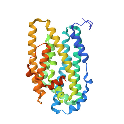
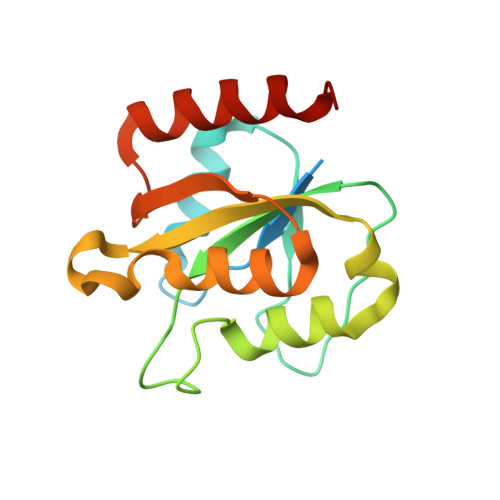
Structural basis for activation of class Ib ribonucleotide reductase.

Boal, A.K.Cotruvo, J.A.Stubbe, J.Rosenzweig, A.C.

(2010) Science 329: 1526-1530

  • DOI: https://doi.org/10.1126/science.1190187
  • Primary Citation Related Structures: 
    3N37, 3N38, 3N39, 3N3A, 3N3B

  • PubMed Abstract: 

    The class Ib ribonucleotide reductase of Escherichia coli can initiate reduction of nucleotides to deoxynucleotides with either a Mn(III)2-tyrosyl radical (Y•) or a Fe(III)2-Y• cofactor in the NrdF subunit. Whereas Fe(III)2-Y• can self-assemble from Fe(II)2-NrdF and O2, activation of Mn(II)2-NrdF requires a reduced flavoprotein, NrdI, proposed to form the oxidant for cofactor assembly by reduction of O2. The crystal structures reported here of E. coli Mn(II)2-NrdF and Fe(II)2-NrdF reveal different coordination environments, suggesting distinct initial binding sites for the oxidants during cofactor activation. In the structures of Mn(II)2-NrdF in complex with reduced and oxidized NrdI, a continuous channel connects the NrdI flavin cofactor to the NrdF Mn(II)2 active site. Crystallographic detection of a putative peroxide in this channel supports the proposed mechanism of Mn(III)2-Y• cofactor assembly.


  • Organizational Affiliation
    • Department of Biochemistry, Molecular Biology and Cell Biology, Northwestern University, Evanston, IL 60208, USA.

Macromolecule Content 

  • Total Structure Weight: 108.58 kDa 
  • Atom Count: 6,885 
  • Modeled Residue Count: 826 
  • Deposited Residue Count: 944 
  • Unique protein chains: 2

Macromolecules

Find similar proteins by:|  3D Structure
Entity ID: 1
MoleculeChains  Sequence LengthOrganismDetailsImage
Ribonucleoside-diphosphate reductase 2 subunit betaA [auth B],
C [auth A]
319Escherichia coli K-12Mutation(s): 0 
Gene Names: b2676JW2651nrdFygaD
EC: 1.17.4.1
UniProt
Find proteins for P37146 (Escherichia coli (strain K12))
Explore P37146 
Go to UniProtKB:  P37146
Entity Groups
Sequence Clusters30% Identity50% Identity70% Identity90% Identity95% Identity100% Identity
UniProt GroupP37146
Sequence Annotations
Expand
Reference Sequence
Find similar proteins by:|  3D Structure
Entity ID: 2
MoleculeChains  Sequence LengthOrganismDetailsImage
Protein nrdIB [auth C],
D
153Escherichia coli K-12Mutation(s): 0 
Gene Names: nrdIygaOb2674JW2649
UniProt
Find proteins for P0A772 (Escherichia coli (strain K12))
Explore P0A772 
Go to UniProtKB:  P0A772
Entity Groups
Sequence Clusters30% Identity50% Identity70% Identity90% Identity95% Identity100% Identity
UniProt GroupP0A772
Sequence Annotations
Expand
Reference Sequence

Small Molecules

Ligands 3 Unique
IDChains Name / Formula / InChI Key2D Diagram3D Interactions
FMN

Query on FMN


Download:Ideal Coordinates CCD File
H [auth C],
L [auth D]
FLAVIN MONONUCLEOTIDE
C17 H21 N4 O9 P
FVTCRASFADXXNN-SCRDCRAPSA-N
MN

Query on MN


Download:Ideal Coordinates CCD File
E [auth B],
F [auth B],
I [auth A],
J [auth A]
MANGANESE (II) ION
Mn
WAEMQWOKJMHJLA-UHFFFAOYSA-N
PEO

Query on PEO


Download:Ideal Coordinates CCD File
G [auth B],
K [auth A]
HYDROGEN PEROXIDE
H2 O2
MHAJPDPJQMAIIY-UHFFFAOYSA-N

Experimental Data & Validation

Experimental Data

  • Method: X-RAY DIFFRACTION
  • Resolution: 2.36 Å
  • R-Value Free:  0.275 (Depositor), 0.263 (DCC) 
  • R-Value Work:  0.234 (Depositor) 
  • R-Value Observed: 0.236 (Depositor) 
Space Group: P 21 21 21
Unit Cell:
Length ( Å )Angle ( ˚ )
a = 75.723α = 90
b = 91.348β = 90
c = 144.114γ = 90
Software Package:
Software NamePurpose
REFMACrefinement
PDB_EXTRACTdata extraction

Structure Validation

View Full Validation Report


Ligand Structure Quality Assessment 


Entry History 

Deposition Data

Revision History  (Full details and data files)

  • Version 1.0: 2010-08-18
    Type: Initial release
  • Version 1.1: 2011-07-13
    Changes: Version format compliance
  • Version 1.2: 2023-09-06
    Changes: Data collection, Database references, Derived calculations, Refinement description