3N00 | pdb_00003n00

Crystal Structure of a deletion mutant of human Reverba ligand binding domain bound with an NCoR ID1 peptide determined to 2.60A


Experimental Data Snapshot

  • Method: X-RAY DIFFRACTION
  • Resolution: 2.60 Å
  • R-Value Free: 
    0.266 (Depositor), 0.270 (DCC) 
  • R-Value Work: 
    0.199 (Depositor), 0.203 (DCC) 
  • R-Value Observed: 
    0.200 (Depositor) 

Starting Model: experimental
View more details

wwPDB Validation 3D Report Full Report

Validation slider image for 3N00

This is version 1.3 of the entry. See complete history

Literature

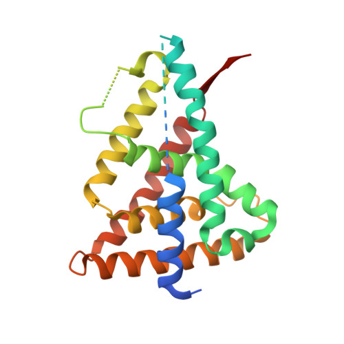
Structure of Rev-erbalpha bound to N-CoR reveals a unique mechanism of nuclear receptor-co-repressor interaction.

Phelan, C.A.Gampe, R.T.Lambert, M.H.Parks, D.J.Montana, V.Bynum, J.Broderick, T.M.Hu, X.Williams, S.P.Nolte, R.T.Lazar, M.A.

(2010) Nat Struct Mol Biol 17: 808-814

  • DOI: https://doi.org/10.1038/nsmb.1860
  • Primary Citation Related Structures: 
    3N00

  • PubMed Abstract: 

    Repression of gene transcription by the nuclear receptor Rev-erbalpha plays an integral role in the core molecular circadian clock. We report the crystal structure of a nuclear receptor-co-repressor (N-CoR) interaction domain 1 (ID1) peptide bound to truncated human Rev-erbalpha ligand-binding domain (LBD). The ID1 peptide forms an unprecedented antiparallel beta-sheet with Rev-erbalpha, as well as an alpha-helix similar to that seen in nuclear receptor ID2 crystal structures but out of register by four residues. Comparison with the structure of Rev-erbbeta bound to heme indicates that ID1 peptide and heme induce substantially different conformational changes in the LBD. Although heme is involved in Rev-erb repression, the structure suggests that Rev-erbalpha could also mediate repression via ID1 binding in the absence of heme. The previously uncharacterized secondary structure induced by ID1 peptide binding advances our understanding of nuclear receptor-co-repressor interactions.


  • Organizational Affiliation
    • Division of Endocrinology, Diabetes and Metabolism, Departments of Medicine and Genetics, The Institute for Diabetes, Obesity and Metabolism, University of Pennsylvania School of Medicine, Philadelphia, Pennsylvania, USA.

Macromolecule Content 

  • Total Structure Weight: 30.08 kDa 
  • Atom Count: 1,611 
  • Modeled Residue Count: 205 
  • Deposited Residue Count: 266 
  • Unique protein chains: 2

Macromolecules

Find similar proteins by:|  3D Structure
Entity ID: 1
MoleculeChains  Sequence LengthOrganismDetailsImage
Rev-erbA-alpha245Homo sapiensMutation(s): 0 
Gene Names: NR1D1EAR1HREVTHRAL
UniProt & NIH Common Fund Data Resources
Find proteins for P20393 (Homo sapiens)
Explore P20393 
Go to UniProtKB:  P20393
PHAROS:  P20393
GTEx:  ENSG00000126368 
Entity Groups
Sequence Clusters30% Identity50% Identity70% Identity90% Identity95% Identity100% Identity
UniProt GroupP20393
Sequence Annotations
Expand
Reference Sequence
Find similar proteins by:  Sequence   |   3D Structure  
Entity ID: 2
MoleculeChains  Sequence LengthOrganismDetailsImage
Nuclear receptor corepressor 121Homo sapiensMutation(s): 0 
UniProt & NIH Common Fund Data Resources
Find proteins for O75376 (Homo sapiens)
Explore O75376 
Go to UniProtKB:  O75376
PHAROS:  O75376
GTEx:  ENSG00000141027 
Entity Groups
Sequence Clusters30% Identity50% Identity70% Identity90% Identity95% Identity100% Identity
UniProt GroupO75376
Sequence Annotations
Expand
Reference Sequence

Experimental Data & Validation

Experimental Data

  • Method: X-RAY DIFFRACTION
  • Resolution: 2.60 Å
  • R-Value Free:  0.266 (Depositor), 0.270 (DCC) 
  • R-Value Work:  0.199 (Depositor), 0.203 (DCC) 
  • R-Value Observed: 0.200 (Depositor) 
Space Group: H 3 2
Unit Cell:
Length ( Å )Angle ( ˚ )
a = 112.55α = 90
b = 112.55β = 90
c = 103.833γ = 120
Software Package:
Software NamePurpose
JDirectordata collection
AMoREphasing
REFMACrefinement
HKL-2000data reduction
HKL-2000data scaling

Structure Validation

View Full Validation Report


Entry History 

Deposition Data

Revision History  (Full details and data files)

  • Version 1.0: 2010-06-30
    Type: Initial release
  • Version 1.1: 2011-07-13
    Changes: Version format compliance
  • Version 1.2: 2017-08-16
    Changes: Refinement description, Source and taxonomy
  • Version 1.3: 2023-09-06
    Changes: Data collection, Database references, Refinement description