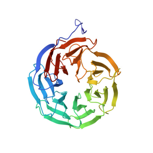
The Effect of Asp-His-Ser/Thr-Trp Tetrad on the Thermostability of WD40-Repeat Proteins
Wu, X.-H., Chen, R.-C., Gao, Y., Wu, Y.-D.(2010) Biochemistry 49: 10237-10245
- PubMed: 20939513
- DOI: https://doi.org/10.1021/bi101321y
- Primary Citation of Related Structures:
3MXX, 3N0D, 3N0E - PubMed Abstract:
We recently found that Asp-His-Ser/Thr-Trp hydrogen-bonded tetrads are widely and uniquely present in the WD40-repeat proteins. WDR5 protein is a seven WD40-repeat propeller with five such tetrads. To explore the effect of the tetrad on the structure and stability of WD40-repeat proteins, the wild-type WDR5 and its seven mutants involving the substitutions of tetrad residues have been isolated. The crystal structures of the wild-type WDR5 and its three WDR5 mutants have been determined by X-ray diffraction method. The mutations of the tetrad residues are found not to change the basic structural features. The denaturing profiles of the wild type and the seven mutants with the use of denaturant guanidine hydrochloride have been studied by circular dichroism spectroscopy to determine the folding free energies of these proteins. The folding free energies of the wild type and the S62A, S146A, S188A, D192E, W330F, W330Y, and D324E mutants are measured to be about -11.6, -2.7, -3.1, -2.9, -3.6, -7.1, -7.0, and -7.5 kcal/mol, respectively. These suggest that (1) the hydrogen bonds in these hydrogen bond networks are unusually strong; (2) each hydrogen-bonded tetrad provides over 12 kcal/mol stability to the protein; thus, the removal of any single tetrad would cause unfolding of the protein; (3) since there are five tetrads, the protein must be in a highly unstable state without the tetrads, which might be related to its biological functions.
- School of Chemical Biology and Biotechnology, Laboratory of Chemical Genomics, Shenzhen Graduate School of Peking University, Shenzhen 518055, China.
Organizational Affiliation:


