Coumarinyl-substituted sulfonamides strongly inhibit several human carbonic anhydrase isoforms: solution and crystallographic investigations.
Wagner, J., Avvaru, B.S., Robbins, A.H., Scozzafava, A., Supuran, C.T., McKenna, R.(2010) Bioorg Med Chem 18: 4873-4878
- PubMed: 20598552 Search on PubMedSearch on PubMed Central
- DOI: https://doi.org/10.1016/j.bmc.2010.06.028
- Primary Citation Related Structures:
3ML2 - PubMed Abstract:
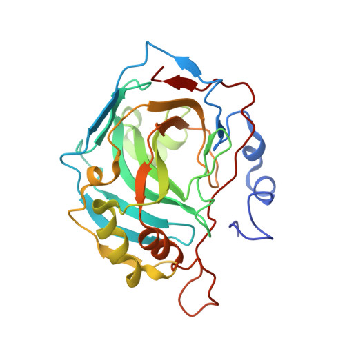
We investigated a series of coumarinyl-substituted aromatic sulfonamides as inhibitors of four carbonic anhydrase (CA, EC 4.2.1.1) isoforms with medical applications, the cytosolic hCA I, and II, and the transmembrane, tumor-associated hCA IX and XII. Compounds incorporating 7-methoxy-coumarin-4-yl-acetamide-tails and benzenesulfonamide and benzene-1,3-disulfonamide scaffolds showed medium potency inhibition of hCA I (KIs of 73-131 nM), effective hCA II inhibition (KIs of 9.1-36 nM) and less effective hCA IX and XII inhibition (KIs of 55-128 nM). Only one compound, the derivatized 4-amino-6-trifluoromethyl-benzene-1,3-disulfonamide with the coumarinyl tail, showed effective inhibition of the transmembrane isoforms, with KIs of 5.9-14.2 nM, although it was less effective as hCA I and II inhibitor (KIs of 36-120 nM). An X-ray crystal structure of hCA II in complex with 4-(7-methoxy-coumarin-4-yl-acetamido)-benzenesulfonamide (KI of 9.1 nM against hCA II) showed the intact inhibitor coordinated to the zinc ion from the enzyme active site by the sulfonamide moiety, and participating in a edge-to-face stacking with Phe131, in addition to other hydrophobic and hydrophilic interactions with water molecules and amino acid residues from the active site. Thus, sulfonamides incorporating coumarin rings have a distinct inhibition mechanism compared to the coumarins, and may lead to compounds with interesting inhibition profiles against various alpha-CAs found in mammals or parasites, such as Plasmodium falciparum.
- Department of Biochemistry and Molecular Biology, College of Medicine, University of Florida, Box 100245, Gainesville, FL 32610, USA.
Organizational Affiliation: