3MKY

Structure of SopB(155-323)-18mer DNA complex, I23 form


Experimental Data Snapshot

  • Method: X-RAY DIFFRACTION
  • Resolution: 2.86 Å
  • R-Value Free: 0.266 
  • R-Value Work: 0.242 
  • R-Value Observed: 0.242 

wwPDB Validation   3D Report Full Report


This is version 1.3 of the entry. See complete history


Literature

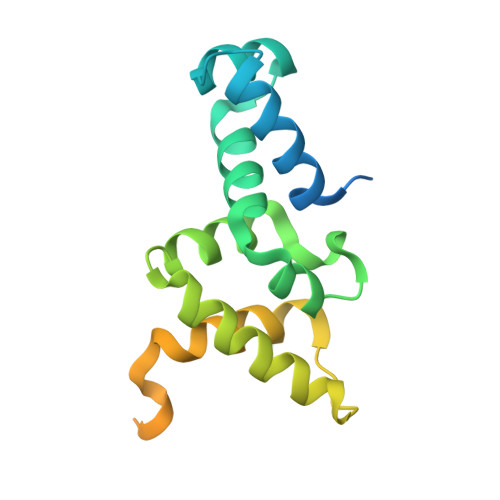
Insight into F plasmid DNA segregation revealed by structures of SopB and SopB-DNA complexes.

Schumacher, M.A.Piro, K.M.Xu, W.

(2010) Nucleic Acids Res 38: 4514-4526

  • DOI: https://doi.org/10.1093/nar/gkq161
  • Primary Citation of Related Structures:  
    3KZ5, 3MKW, 3MKY, 3MKZ

  • PubMed Abstract: 

    Accurate DNA segregation is essential for genome transmission. Segregation of the prototypical F plasmid requires the centromere-binding protein SopB, the NTPase SopA and the sopC centromere. SopB displays an intriguing range of DNA-binding properties essential for partition; it binds sopC to form a partition complex, which recruits SopA, and it also coats DNA to prevent non-specific SopA-DNA interactions, which inhibits SopA polymerization. To understand the myriad functions of SopB, we determined a series of SopB-DNA crystal structures. SopB does not distort its DNA site and our data suggest that SopB-sopC forms an extended rather than wrapped partition complex with the SopA-interacting domains aligned on one face. SopB is a multidomain protein, which like P1 ParB contains an all-helical DNA-binding domain that is flexibly attached to a compact (beta(3)-alpha)(2) dimer-domain. Unlike P1 ParB, the SopB dimer-domain does not bind DNA. Moreover, SopB contains a unique secondary dimerization motif that bridges between DNA duplexes. Both specific and non-specific SopB-DNA bridging structures were observed. This DNA-linking function suggests a novel mechanism for in trans DNA spreading by SopB, explaining how it might mask DNA to prevent DNA-mediated inhibition of SopA polymerization.


  • Organizational Affiliation

    Department of Biochemistry and Molecular Biology, University of Texas, M.D. Anderson Cancer Center, Unit 1000, Houston, TX 77030, USA. maschuma@mdanderson.org


Macromolecules

Find similar proteins by:  (by identity cutoff)  |  3D Structure
Entity ID: 2
MoleculeChains Sequence LengthOrganismDetailsImage
Protein sopBC [auth B],
D [auth P]
189Escherichia coli K-12Mutation(s): 0 
Gene Names: BECOK12F047plasmidsopB
UniProt
Find proteins for P62558 (Escherichia coli (strain K12))
Explore P62558 
Go to UniProtKB:  P62558
Entity Groups  
Sequence Clusters30% Identity50% Identity70% Identity90% Identity95% Identity100% Identity
UniProt GroupP62558
Sequence Annotations
Expand
  • Reference Sequence

Find similar nucleic acids by:  Sequence   |   3D Structure  

Entity ID: 1
MoleculeChains LengthOrganismImage
DNA (5'-D(*CP*TP*GP*GP*GP*AP*CP*CP*AP*TP*GP*GP*TP*CP*CP*CP*AP*G)-3')A [auth U],
B [auth T]
18N/A
Sequence Annotations
Expand
  • Reference Sequence
Experimental Data & Validation

Experimental Data

  • Method: X-RAY DIFFRACTION
  • Resolution: 2.86 Å
  • R-Value Free: 0.266 
  • R-Value Work: 0.242 
  • R-Value Observed: 0.242 
  • Space Group: I 2 3
Unit Cell:
Length ( Å )Angle ( ˚ )
a = 200.78α = 90
b = 200.78β = 90
c = 200.78γ = 90
Software Package:
Software NamePurpose
ADSCdata collection
SOLVEphasing
CNSrefinement
MOSFLMdata reduction
SCALAdata scaling

Structure Validation

View Full Validation Report


Entry History 

Deposition Data

Revision History  (Full details and data files)

  • Version 1.0: 2010-05-05
    Type: Initial release
  • Version 1.1: 2011-07-13
    Changes: Version format compliance
  • Version 1.2: 2015-01-21
    Changes: Database references, Structure summary
  • Version 1.3: 2024-02-21
    Changes: Data collection, Database references, Derived calculations