3M3X | pdb_00003m3x

Crystal structure of human carbonic anhydrase isozyme II with 4-{2-[N-(6-methoxy-5-nitropyrimidin-4-yl)amino]ethyl}benzenesulfonamide


Experimental Data Snapshot

  • Method: X-RAY DIFFRACTION
  • Resolution: 1.68 Å
  • R-Value Free: 
    0.222 (Depositor), 0.219 (DCC) 
  • R-Value Work: 
    0.163 (Depositor), 0.159 (DCC) 
  • R-Value Observed: 
    0.168 (Depositor) 

Starting Model: experimental
View more details

wwPDB Validation 3D Report Full Report

Validation slider image for 3M3X

Ligand Structure Quality Assessment 


This is version 1.3 of the entry. See complete history

Literature

4-[N-(Substituted 4-pyrimidinyl)amino]benzenesulfonamides as inhibitors of carbonic anhydrase isozymes I, II, VII, and XIII

Sudzius, J.Baranauskiene, L.Golovenko, D.Matuliene, J.Michailoviene, V.Torresan, J.Jachno, J.Sukackaite, R.Manakova, E.Grazulis, S.Tumkevicius, S.Matulis, D.

(2010) Bioorg Med Chem 18: 7413-7421

  • DOI: https://doi.org/10.1016/j.bmc.2010.09.011
  • Primary Citation Related Structures: 
    3M3X, 3M40, 3M5E, 3MHI, 3MHL, 3MHM, 3MHO

  • PubMed Abstract: 

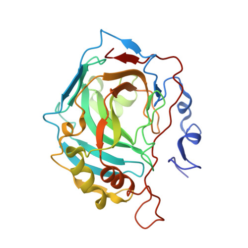
    A series of 4-[N-(substituted 4-pyrimidinyl)amino]benzenesulfonamides were designed and synthesised. Their binding potencies as inhibitors of selected recombinant human carbonic anhydrase (hCA) isozymes I, II, VII, and XIII were measured using isothermal titration calorimetry and the thermal shift assay. To determine the structural features of inhibitor binding, the crystal structures of several compounds in complex with hCA II were determined. Several compounds exhibited selectivity towards isozymes I, II, and XIII, and some were potent inhibitors of hCA VII.


  • Organizational Affiliation
    • Department of Organic Chemistry, Faculty of Chemistry, Vilnius University, Naugarduko 24, LT-03225 Vilnius, Lithuania.

Macromolecule Content 

  • Total Structure Weight: 29.79 kDa 
  • Atom Count: 2,507 
  • Modeled Residue Count: 258 
  • Deposited Residue Count: 260 
  • Unique protein chains: 1

Macromolecules

Find similar proteins by:|  3D Structure
Entity ID: 1
MoleculeChains  Sequence LengthOrganismDetailsImage
Carbonic anhydrase 2260Homo sapiensMutation(s): 0 
EC: 4.2.1.1 (PDB Primary Data), 4.2.1.69 (UniProt)
UniProt & NIH Common Fund Data Resources
Find proteins for P00918 (Homo sapiens)
Explore P00918 
Go to UniProtKB:  P00918
PHAROS:  P00918
GTEx:  ENSG00000104267 
Entity Groups
Sequence Clusters30% Identity50% Identity70% Identity90% Identity95% Identity100% Identity
UniProt GroupP00918
Sequence Annotations
Expand
Reference Sequence

Experimental Data & Validation

Experimental Data

  • Method: X-RAY DIFFRACTION
  • Resolution: 1.68 Å
  • R-Value Free:  0.222 (Depositor), 0.219 (DCC) 
  • R-Value Work:  0.163 (Depositor), 0.159 (DCC) 
  • R-Value Observed: 0.168 (Depositor) 
Space Group: P 1 21 1
Unit Cell:
Length ( Å )Angle ( ˚ )
a = 42.035α = 90
b = 41.051β = 104.39
c = 71.805γ = 90
Software Package:
Software NamePurpose
REFMACrefinement
MOSFLMdata reduction
SCALAdata scaling
PDB_EXTRACTdata extraction
MAR345data collection
Omodel building

Structure Validation

View Full Validation Report


Ligand Structure Quality Assessment 


Entry History 

Deposition Data

Revision History  (Full details and data files)

  • Version 1.0: 2010-10-20
    Type: Initial release
  • Version 1.1: 2011-07-13
    Changes: Version format compliance
  • Version 1.2: 2017-11-08
    Changes: Advisory, Refinement description
  • Version 1.3: 2023-11-01
    Changes: Advisory, Data collection, Database references, Derived calculations, Refinement description