3LSL

Piracetam bound to the ligand binding domain of GluA2 (flop form)


Experimental Data Snapshot

  • Method: X-RAY DIFFRACTION
  • Resolution: 2.12 Å
  • R-Value Free: 0.263 
  • R-Value Work: 0.201 
  • R-Value Observed: 0.203 

wwPDB Validation   3D Report Full Report


This is version 1.3 of the entry. See complete history


Literature

Piracetam Defines a New Binding Site for Allosteric Modulators of alpha-Amino-3-hydroxy-5-methyl-4-isoxazole-propionic Acid (AMPA) Receptors.

Ahmed, A.H.Oswald, R.E.

(2010) J Med Chem 53: 2197-2203

  • DOI: https://doi.org/10.1021/jm901905j
  • Primary Citation of Related Structures:  
    3LSF, 3LSL, 3LSW, 3LSX

  • PubMed Abstract: 

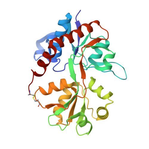
    Glutamate receptors are the most prevalent excitatory neurotransmitter receptors in the vertebrate central nervous system and are important potential drug targets for cognitive enhancement and the treatment of schizophrenia. Allosteric modulators of AMPA receptors promote dimerization by binding to a dimer interface and reducing desensitization and deactivation. The pyrrolidine allosteric modulators, piracetam and aniracetam, were among the first of this class of drugs to be discovered. We have determined the structure of the ligand binding domain of the AMPA receptor subtypes GluA2 and GluA3 with piracetam and a corresponding structure of GluA3 with aniracetam. Both drugs bind to GluA2 and GluA3 in a very similar manner, suggesting little subunit specificity. However, the binding sites for piracetam and aniracetam differ considerably. Aniracetam binds to a symmetrical site at the center of the dimer interface. Piracetam binds to multiple sites along the dimer interface with low occupation, one of which is a unique binding site for potential allosteric modulators. This new site may be of importance in the design of new allosteric regulators.


  • Organizational Affiliation

    Department of Molecular Medicine, Cornell University, Ithaca, New York 14853, USA.


Macromolecules
Find similar proteins by:  (by identity cutoff)  |  3D Structure
Entity ID: 1
MoleculeChains Sequence LengthOrganismDetailsImage
Glutamate receptor 2A,
B [auth D],
C [auth G]
258Rattus norvegicusMutation(s): 0 
Gene Names: Gria2Glur2
UniProt
Find proteins for P19491 (Rattus norvegicus)
Explore P19491 
Go to UniProtKB:  P19491
Entity Groups  
Sequence Clusters30% Identity50% Identity70% Identity90% Identity95% Identity100% Identity
UniProt GroupP19491
Sequence Annotations
Expand
  • Reference Sequence
Small Molecules
Ligands 3 Unique
IDChains Name / Formula / InChI Key2D Diagram3D Interactions
GLU
Query on GLU

Download Ideal Coordinates CCD File 
D [auth A],
J [auth D],
P [auth G]
GLUTAMIC ACID
C5 H9 N O4
WHUUTDBJXJRKMK-VKHMYHEASA-N
PZI
Query on PZI

Download Ideal Coordinates CCD File 
E [auth A]
F [auth A]
G [auth A]
K [auth D]
L [auth D]
E [auth A],
F [auth A],
G [auth A],
K [auth D],
L [auth D],
M [auth D],
Q [auth G],
R [auth G],
S [auth G]
2-(2-oxopyrrolidin-1-yl)acetamide
C6 H10 N2 O2
GMZVRMREEHBGGF-UHFFFAOYSA-N
ZN
Query on ZN

Download Ideal Coordinates CCD File 
H [auth A],
I [auth A],
N [auth D],
O [auth D],
T [auth G]
ZINC ION
Zn
PTFCDOFLOPIGGS-UHFFFAOYSA-N
Experimental Data & Validation

Experimental Data

  • Method: X-RAY DIFFRACTION
  • Resolution: 2.12 Å
  • R-Value Free: 0.263 
  • R-Value Work: 0.201 
  • R-Value Observed: 0.203 
  • Space Group: P 2 21 21
Unit Cell:
Length ( Å )Angle ( ˚ )
a = 47.528α = 90
b = 114.343β = 90
c = 163.829γ = 90
Software Package:
Software NamePurpose
HKL-2000data collection
PHENIXmodel building
PHENIXrefinement
HKL-2000data reduction
HKL-2000data scaling
PHENIXphasing

Structure Validation

View Full Validation Report


Entry History 

Deposition Data

Revision History  (Full details and data files)

  • Version 1.0: 2010-03-16
    Type: Initial release
  • Version 1.1: 2011-07-13
    Changes: Version format compliance
  • Version 1.2: 2017-08-02
    Changes: Source and taxonomy
  • Version 1.3: 2023-09-06
    Changes: Data collection, Database references, Derived calculations, Refinement description