Systematic mutational analysis of peptide inhibition of the p53-MDM2/MDMX interactions.
Li, C., Pazgier, M., Li, C., Yuan, W., Liu, M., Wei, G., Lu, W.Y., Lu, W.(2010) J Mol Biol 398: 200-213
- PubMed: 20226197 Search on PubMedSearch on PubMed Central
- DOI: https://doi.org/10.1016/j.jmb.2010.03.005
- Primary Citation Related Structures:
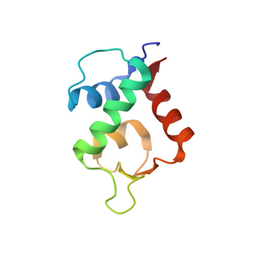
3LNZ - PubMed Abstract:
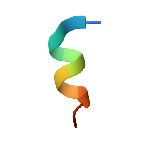
Inhibition of the interaction between the tumor suppressor protein p53 and its negative regulators MDM2 and MDMX is of great interest in cancer biology and drug design. We previously reported a potent duodecimal peptide inhibitor, termed PMI (TSFAEYWNLLSP), of the p53-MDM2 and -MDMX interactions. PMI competes with p53 for MDM2 and MDMX binding at an affinity roughly 2 orders of magnitude higher than that of (17-28)p53 (ETFSDLWKLLPE) of the same length; both peptides adopt nearly identical alpha-helical conformations in the complexes, where the three highlighted hydrophobic residues Phe, Trp, and Leu dominate PMI or (17-28)p53 binding to MDM2 and MDMX. To elucidate the molecular determinants for PMI activity and specificity, we performed a systematic Ala scanning mutational analysis of PMI and (17-28)p53. The binding affinities for MDM2 and MDMX of a total of 35 peptides including 10 truncation analogs were quantified, affording a complete dissection of energetic contributions of individual residues of PMI and (17-28)p53 to MDM2 and MDMX association. Importantly, the N8A mutation turned PMI into the most potent dual-specific antagonist of MDM2 and MDMX reported to date, registering respective K(d) values of 490 pM and 2.4 nM. The co-crystal structure of N8A-PMI-(25-109)MDM2 was determined at 1.95 A, affirming that high-affinity peptide binding to MDM2/MDMX necessitates, in addition to optimized intermolecular interactions, enhanced helix stability or propensity contributed by non-contact residues. The powerful empirical binding data and crystal structures present a unique opportunity for computational studies of peptide inhibition of the p53-MDM2/MDMX interactions.
- Institute of Human Virology, University of Maryland School of Medicine, 725 West Lombard Street, Baltimore, MD 21201, USA.
Organizational Affiliation: