3LCP | pdb_00003lcp

Crystal structure of the carbohydrate recognition domain of LMAN1 in complex with MCFD2


Experimental Data Snapshot

  • Method: X-RAY DIFFRACTION
  • Resolution: 2.45 Å
  • R-Value Free: 
    0.247 (Depositor), 0.233 (DCC) 
  • R-Value Work: 
    0.196 (Depositor), 0.195 (DCC) 
  • R-Value Observed: 
    0.199 (Depositor) 

Starting Model: experimental
View more details

wwPDB Validation 3D Report Full Report

Validation slider image for 3LCP

This is version 1.3 of the entry. See complete history

Literature

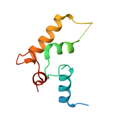
Crystal structure of the LMAN1-CRD/MCFD2 transport receptor complex provides insight into combined deficiency of factor V and factor VIII.

Wigren, E.Bourhis, J.M.Kursula, I.Guy, J.E.Lindqvist, Y.

(2010) FEBS Lett 584: 878-882

  • DOI: https://doi.org/10.1016/j.febslet.2010.02.009
  • Primary Citation Related Structures: 
    3LCP

  • PubMed Abstract: 

    LMAN1 is a glycoprotein receptor, mediating transfer from the ER to the ER-Golgi intermediate compartment. Together with the co-receptor MCFD2, it transports coagulation factors V and VIII. Mutations in LMAN1 and MCFD2 can cause combined deficiency of factors V and VIII (F5F8D). We present the crystal structure of the LMAN1/MCFD2 complex and relate it to patient mutations. Circular dichroism data show that the majority of the substitution mutations give rise to a disordered or severely destabilized MCFD2 protein. The few stable mutation variants are found in the binding surface of the complex leading to impaired LMAN1 binding and F5F8D.


  • Organizational Affiliation
    • Department of Medical Biochemistry and Biophysics, Karolinska Institutet, 17177 Stockholm, Sweden.

Macromolecule Content 

  • Total Structure Weight: 75.51 kDa 
  • Atom Count: 4,888 
  • Modeled Residue Count: 605 
  • Deposited Residue Count: 680 
  • Unique protein chains: 2

Macromolecules

Find similar proteins by:|  3D Structure
Entity ID: 1
MoleculeChains  Sequence LengthOrganismDetailsImage
Protein ERGIC-53
A, B
247Homo sapiensMutation(s): 0 
Gene Names: ERGIC53F5F8DLMAN1LMAN1 (AMINO ACIDS 32-277)
UniProt & NIH Common Fund Data Resources
Find proteins for P49257 (Homo sapiens)
Explore P49257 
Go to UniProtKB:  P49257
PHAROS:  P49257
GTEx:  ENSG00000074695 
Entity Groups
Sequence Clusters30% Identity50% Identity70% Identity90% Identity95% Identity100% Identity
UniProt GroupP49257
Sequence Annotations
Expand
Reference Sequence
Find similar proteins by:|  3D Structure
Entity ID: 2
MoleculeChains  Sequence LengthOrganismDetailsImage
Multiple coagulation factor deficiency protein 2
C, D
93Homo sapiensMutation(s): 0 
Gene Names: MCFD2MCFD2 (AMINO ACIDS 58-146)SDNSF
UniProt & NIH Common Fund Data Resources
Find proteins for Q8NI22 (Homo sapiens)
Explore Q8NI22 
Go to UniProtKB:  Q8NI22
PHAROS:  Q8NI22
GTEx:  ENSG00000180398 
Entity Groups
Sequence Clusters30% Identity50% Identity70% Identity90% Identity95% Identity100% Identity
UniProt GroupQ8NI22
Sequence Annotations
Expand
Reference Sequence

Experimental Data & Validation

Experimental Data

  • Method: X-RAY DIFFRACTION
  • Resolution: 2.45 Å
  • R-Value Free:  0.247 (Depositor), 0.233 (DCC) 
  • R-Value Work:  0.196 (Depositor), 0.195 (DCC) 
  • R-Value Observed: 0.199 (Depositor) 
Space Group: P 61
Unit Cell:
Length ( Å )Angle ( ˚ )
a = 58.602α = 90
b = 58.602β = 90
c = 396.87γ = 120
Software Package:
Software NamePurpose
DNAdata collection
PHASERphasing
REFMACrefinement
XDSdata reduction
SCALAdata scaling

Structure Validation

View Full Validation Report


Entry History 

Deposition Data

Revision History  (Full details and data files)

  • Version 1.0: 2010-01-26
    Type: Initial release
  • Version 1.1: 2011-07-13
    Changes: Version format compliance
  • Version 1.2: 2023-09-06
    Changes: Data collection, Database references, Derived calculations, Refinement description
  • Version 1.3: 2024-11-06
    Changes: Structure summary