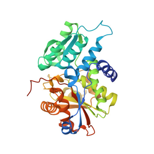
3L6C

X-ray crystal structure of rat serine racemase in complex with malonate a potent inhibitor


Experimental Data Snapshot

  • Method: X-RAY DIFFRACTION
  • Resolution: 2.20 Å
  • R-Value Free: 0.267 
  • R-Value Work: 0.216 
  • R-Value Observed: 0.219 

wwPDB Validation   3D Report Full Report


Ligand Structure Quality Assessment 


This is version 1.3 of the entry. See complete history


Literature

The structure of mammalian serine racemase: evidence for conformational changes upon inhibitor binding.

Smith, M.A.Mack, V.Ebneth, A.Moraes, I.Felicetti, B.Wood, M.Schonfeld, D.Mather, O.Cesura, A.Barker, J.

(2010) J Biol Chem 285: 12873-12881

  • DOI: https://doi.org/10.1074/jbc.M109.050062
  • Primary Citation of Related Structures:  
    3HMK, 3L6B, 3L6C, 3L6R

  • PubMed Abstract: 

    Serine racemase is responsible for the synthesis of D-serine, an endogenous co-agonist for N-methyl-D-aspartate receptor-type glutamate receptors (NMDARs). This pyridoxal 5'-phosphate-dependent enzyme is involved both in the reversible conversion of L- to D-serine and serine catabolism by alpha,beta-elimination of water, thereby regulating D-serine levels. Because D-serine affects NMDAR signaling throughout the brain, serine racemase is a promising target for the treatment of disorders related to NMDAR dysfunction. To provide a molecular basis for rational drug design the x-ray crystal structures of human and rat serine racemase were determined at 1.5- and 2.1-A resolution, respectively, and in the presence and absence of the orthosteric inhibitor malonate. The structures revealed a fold typical of beta-family pyridoxal 5'-phosphate enzymes, with both a large domain and a flexible small domain associated into a symmetric dimer, and indicated a ligand-induced rearrangement of the small domain that organizes the active site for specific turnover of the substrate.


  • Organizational Affiliation

    Department of Structural Biology and Biology, Evotec, 114 Milton Park, Abingdon, Oxon OX14 4SA, United Kingdom. myron.smith@evotec.com


Macromolecules
Find similar proteins by:  (by identity cutoff)  |  3D Structure
Entity ID: 1
MoleculeChains Sequence LengthOrganismDetailsImage
Serine racemase
A, B
339Rattus norvegicusMutation(s): 2 
Gene Names: Srr
EC: 5.1.1.18
UniProt
Find proteins for Q76EQ0 (Rattus norvegicus)
Explore Q76EQ0 
Go to UniProtKB:  Q76EQ0
Entity Groups  
Sequence Clusters30% Identity50% Identity70% Identity90% Identity95% Identity100% Identity
UniProt GroupQ76EQ0
Sequence Annotations
Expand
  • Reference Sequence
Experimental Data & Validation

Experimental Data

  • Method: X-RAY DIFFRACTION
  • Resolution: 2.20 Å
  • R-Value Free: 0.267 
  • R-Value Work: 0.216 
  • R-Value Observed: 0.219 
  • Space Group: P 3
Unit Cell:
Length ( Å )Angle ( ˚ )
a = 110.593α = 90
b = 110.593β = 90
c = 47.917γ = 120
Software Package:
Software NamePurpose
CrystalCleardata collection
PHASERphasing
REFMACrefinement
d*TREKdata reduction
d*TREKdata scaling

Structure Validation

View Full Validation Report


Ligand Structure Quality Assessment 


Entry History 

Deposition Data

Revision History  (Full details and data files)

  • Version 1.0: 2010-01-26
    Type: Initial release
  • Version 1.1: 2011-07-13
    Changes: Version format compliance
  • Version 1.2: 2021-11-10
    Changes: Database references, Derived calculations
  • Version 1.3: 2024-04-03
    Changes: Data collection, Refinement description