3KWW | pdb_00003kww

Crystal structure of the 'restriction triad' mutant of HLA B*3508, beta-2-microglobulin and EBV peptide


Experimental Data Snapshot

  • Method: X-RAY DIFFRACTION
  • Resolution: 2.18 Å
  • R-Value Free: 
    0.251 (Depositor), 0.221 (DCC) 
  • R-Value Work: 
    0.186 (Depositor) 
  • R-Value Observed: 
    0.188 (Depositor) 

Starting Model: experimental
View more details

wwPDB Validation 3D Report Full Report

Validation slider image for 3KWW

This is version 1.4 of the entry. See complete history

Literature

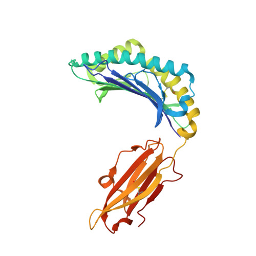
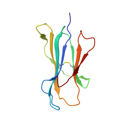
Hard wiring of T cell receptor specificity for the major histocompatibility complex is underpinned by TCR adaptability

Burrows, S.R.Chen, Z.Archbold, J.K.Tynan, F.E.Beddoe, T.Kjer-Nielsen, L.Miles, J.J.Khanna, R.Moss, D.J.Liu, Y.C.Gras, S.Kostenko, L.Brennan, R.M.Clements, C.S.Brooks, A.G.Purcell, A.W.McCluskey, J.Rossjohn, J.

(2010) Proc Natl Acad Sci U S A 

  • DOI: https://doi.org/10.1073/pnas.1004926107
  • Primary Citation Related Structures: 
    3KWW, 3KXF

  • PubMed Abstract: 

    alphabeta T cell receptors (TCRs) are genetically restricted to corecognize peptide antigens bound to self-major histocompatibility complex (pMHC) molecules; however, the basis for this MHC specificity remains unclear. Despite the current dogma, evaluation of the TCR-pMHC-I structural database shows that the nongermline-encoded complementarity-determining region (CDR)-3 loops often contact the MHC-I, and the germline-encoded CDR1 and -2 loops frequently participate in peptide-mediated interactions. Nevertheless, different TCRs adopt a roughly conserved docking mode over the pMHC-I, in which three MHC-I residues (65, 69, and 155) are invariably contacted by the TCR in one way or another. Nonetheless, the impact of mutations at these three positions, either individually or together, was not uniformly detrimental to TCR recognition of pHLA-B*0801 or pHLA-B*3508. Moreover, when TCR-pMHC-I recognition was impaired, this could be partially restored by expression of the CD8 coreceptor. The structure of a TCR-pMHC-I complex in which these three (65, 69, and 155) MHC-I positions were all mutated resulted in shifting of the TCR footprint relative to the cognate complex and formation of compensatory interactions. Collectively, our findings reveal the inherent adaptability of the TCR in maintaining peptide recognition while accommodating changes to the central docking site on the pMHC-I.


  • Organizational Affiliation
    • Cellular Immunology Laboratory, Queensland Institute of Medical Research and Australian Centre for Vaccine Development, Brisbane 4029, Australia.

Macromolecule Content 

  • Total Structure Weight: 46.38 kDa 
  • Atom Count: 3,714 
  • Modeled Residue Count: 388 
  • Deposited Residue Count: 388 
  • Unique protein chains: 3

Macromolecules

Find similar proteins by:|  3D Structure
Entity ID: 1
MoleculeChains  Sequence LengthOrganismDetailsImage
HLA class I histocompatibility antigen, B-35 alpha chain276Homo sapiensMutation(s): 3 
UniProt & NIH Common Fund Data Resources
Find proteins for P01889 (Homo sapiens)
Explore P01889 
Go to UniProtKB:  P01889
PHAROS:  P01889
GTEx:  ENSG00000234745 
Entity Groups
Sequence Clusters30% Identity50% Identity70% Identity90% Identity95% Identity100% Identity
UniProt GroupP01889
Sequence Annotations
Expand
Reference Sequence
Find similar proteins by:|  3D Structure
Entity ID: 2
MoleculeChains  Sequence LengthOrganismDetailsImage
Beta-2-microglobulin99Homo sapiensMutation(s): 0 
UniProt & NIH Common Fund Data Resources
Find proteins for P61769 (Homo sapiens)
Explore P61769 
Go to UniProtKB:  P61769
PHAROS:  P61769
GTEx:  ENSG00000166710 
Entity Groups
Sequence Clusters30% Identity50% Identity70% Identity90% Identity95% Identity100% Identity
UniProt GroupP61769
Sequence Annotations
Expand
Reference Sequence
Find similar proteins by:  Sequence   |   3D Structure  
Entity ID: 3
MoleculeChains  Sequence LengthOrganismDetailsImage
peptide from Trans-activator protein BZLF113N/AMutation(s): 0 
UniProt
Find proteins for P03206 (Epstein-Barr virus (strain B95-8))
Explore P03206 
Go to UniProtKB:  P03206
Entity Groups
Sequence Clusters30% Identity50% Identity70% Identity90% Identity95% Identity100% Identity
UniProt GroupP03206
Sequence Annotations
Expand
Reference Sequence

Small Molecules

Ligands 3 Unique
IDChains Name / Formula / InChI Key2D Diagram3D Interactions
PEG

Query on PEG


Download:Ideal Coordinates CCD File
L [auth A]
M [auth A]
N [auth A]
O [auth A]
P [auth A]
L [auth A],
M [auth A],
N [auth A],
O [auth A],
P [auth A],
Q [auth A],
R [auth B],
S [auth C]
DI(HYDROXYETHYL)ETHER
C4 H10 O3
MTHSVFCYNBDYFN-UHFFFAOYSA-N
GOL

Query on GOL


Download:Ideal Coordinates CCD File
K [auth A]GLYCEROL
C3 H8 O3
PEDCQBHIVMGVHV-UHFFFAOYSA-N
ACY

Query on ACY


Download:Ideal Coordinates CCD File
D [auth A]
E [auth A]
F [auth A]
G [auth A]
H [auth A]
D [auth A],
E [auth A],
F [auth A],
G [auth A],
H [auth A],
I [auth A],
J [auth A]
ACETIC ACID
C2 H4 O2
QTBSBXVTEAMEQO-UHFFFAOYSA-N

Experimental Data & Validation

Experimental Data

  • Method: X-RAY DIFFRACTION
  • Resolution: 2.18 Å
  • R-Value Free:  0.251 (Depositor), 0.221 (DCC) 
  • R-Value Work:  0.186 (Depositor) 
  • R-Value Observed: 0.188 (Depositor) 
Space Group: P 21 21 21
Unit Cell:
Length ( Å )Angle ( ˚ )
a = 50.972α = 90
b = 81.454β = 90
c = 110.38γ = 90
Software Package:
Software NamePurpose
DENZOdata reduction
SCALEPACKdata scaling
PHASERphasing
REFMACrefinement
PDB_EXTRACTdata extraction

Structure Validation

View Full Validation Report


Entry History 

Deposition Data

Revision History  (Full details and data files)

  • Version 1.0: 2010-06-09
    Type: Initial release
  • Version 1.1: 2011-07-13
    Changes: Version format compliance
  • Version 1.2: 2017-11-01
    Changes: Advisory, Refinement description
  • Version 1.3: 2023-11-01
    Changes: Advisory, Data collection, Database references, Derived calculations, Refinement description
  • Version 1.4: 2024-10-30
    Changes: Structure summary