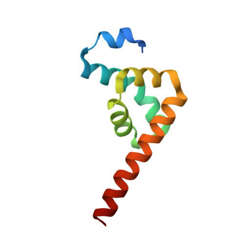
3KUS

Crystal structure of the MLLE domain of poly(A)-binding protein in complex with the binding region of Paip2


Experimental Data Snapshot

  • Method: X-RAY DIFFRACTION
  • Resolution: 1.40 Å
  • R-Value Free: 0.205 
  • R-Value Work: 0.178 
  • R-Value Observed: 0.179 

Starting Model: experimental
View more details

wwPDB Validation   3D Report Full Report


This is version 1.2 of the entry. See complete history


Literature

Molecular Determinants of PAM2 Recognition by the MLLE Domain of Poly(A)-Binding Protein.

Kozlov, G.Menade, M.Rosenauer, A.Nguyen, L.Gehring, K.

(2010) J Mol Biol 397: 397-407

  • DOI: https://doi.org/10.1016/j.jmb.2010.01.032
  • Primary Citation of Related Structures:  
    3KUR, 3KUS, 3KUT

  • PubMed Abstract: 

    MLLE (previously known as PABC) is a peptide-binding domain that is found in poly(A)-binding protein (PABP) and EDD (E3 isolated by differential display), a HECT E3 ubiquitin ligase also known as HYD (hyperplastic discs tumor suppressor) or UBR5. The MLLE domain from PABP recruits various regulatory proteins and translation factors to poly(A) mRNAs through binding of a conserved 12 amino acid peptide motif called PAM2 (for PABP-interacting motif 2). Here, we determined crystal structures of the MLLE domain from PABP alone and in complex with PAM2 peptides from PABP-interacting protein 2. The structures provide a detailed view of hydrophobic determinants of the MLLE binding coded by PAM2 positions 3, 5, 7, 10, and 12 and reveal novel intermolecular polar contacts. In particular, the side chain of the invariant MLLE residue K580 forms hydrogen bonds with the backbone of PAM2 residues 5 and 7. The structures also show that peptide residues outside of the conserved PAM2 motif contribute to binding. Altogether, the structures provide a significant advance in understanding the molecular basis for the binding of PABP by PAM2-containing proteins involved in translational control, mRNA deadenylation, and other cellular processes.


  • Organizational Affiliation

    Department of Biochemistry, Groupe de recherche axé sur la structure des protéines, McGill University, 3655 Promenade Sir William Osler, Montréal, Québec, Canada H3G 1Y6.


Macromolecules
Find similar proteins by:  (by identity cutoff)  |  3D Structure
Entity ID: 1
MoleculeChains Sequence LengthOrganismDetailsImage
Polyadenylate-binding protein 1
A, B
88Homo sapiensMutation(s): 0 
Gene Names: PABPC1PAB1PABP1PABPC2
UniProt & NIH Common Fund Data Resources
Find proteins for P11940 (Homo sapiens)
Explore P11940 
Go to UniProtKB:  P11940
PHAROS:  P11940
GTEx:  ENSG00000070756 
Entity Groups  
Sequence Clusters30% Identity50% Identity70% Identity90% Identity95% Identity100% Identity
UniProt GroupP11940
Sequence Annotations
Expand
  • Reference Sequence

Find similar proteins by:  Sequence   |   3D Structure  

Entity ID: 2
MoleculeChains Sequence LengthOrganismDetailsImage
PAIP2 protein
C, D
15Homo sapiensMutation(s): 0 
UniProt & NIH Common Fund Data Resources
Find proteins for Q9BPZ3 (Homo sapiens)
Explore Q9BPZ3 
Go to UniProtKB:  Q9BPZ3
PHAROS:  Q9BPZ3
GTEx:  ENSG00000120727 
Entity Groups  
Sequence Clusters30% Identity50% Identity70% Identity90% Identity95% Identity100% Identity
UniProt GroupQ9BPZ3
Sequence Annotations
Expand
  • Reference Sequence
Small Molecules
Experimental Data & Validation

Experimental Data

  • Method: X-RAY DIFFRACTION
  • Resolution: 1.40 Å
  • R-Value Free: 0.205 
  • R-Value Work: 0.178 
  • R-Value Observed: 0.179 
  • Space Group: P 1
Unit Cell:
Length ( Å )Angle ( ˚ )
a = 26.396α = 100.12
b = 31.61β = 92.26
c = 48.214γ = 98.9
Software Package:
Software NamePurpose
ADSCdata collection
PHASERphasing
REFMACrefinement
HKL-2000data reduction
HKL-2000data scaling

Structure Validation

View Full Validation Report


Entry History 

Deposition Data

Revision History  (Full details and data files)

  • Version 1.0: 2010-02-09
    Type: Initial release
  • Version 1.1: 2011-07-13
    Changes: Advisory, Version format compliance
  • Version 1.2: 2023-09-06
    Changes: Data collection, Database references, Derived calculations, Refinement description