Molecular Determinants of PAM2 Recognition by the MLLE Domain of Poly(A)-Binding Protein.
Kozlov, G., Menade, M., Rosenauer, A., Nguyen, L., Gehring, K.(2010) J Mol Biol 397: 397-407
- PubMed: 20096703 Search on PubMed
- DOI: https://doi.org/10.1016/j.jmb.2010.01.032
- Primary Citation Related Structures:
3KUR, 3KUS, 3KUT - PubMed Abstract:
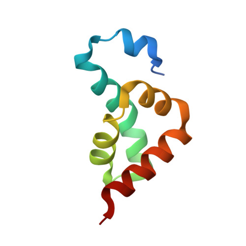
MLLE (previously known as PABC) is a peptide-binding domain that is found in poly(A)-binding protein (PABP) and EDD (E3 isolated by differential display), a HECT E3 ubiquitin ligase also known as HYD (hyperplastic discs tumor suppressor) or UBR5. The MLLE domain from PABP recruits various regulatory proteins and translation factors to poly(A) mRNAs through binding of a conserved 12 amino acid peptide motif called PAM2 (for PABP-interacting motif 2). Here, we determined crystal structures of the MLLE domain from PABP alone and in complex with PAM2 peptides from PABP-interacting protein 2. The structures provide a detailed view of hydrophobic determinants of the MLLE binding coded by PAM2 positions 3, 5, 7, 10, and 12 and reveal novel intermolecular polar contacts. In particular, the side chain of the invariant MLLE residue K580 forms hydrogen bonds with the backbone of PAM2 residues 5 and 7. The structures also show that peptide residues outside of the conserved PAM2 motif contribute to binding. Altogether, the structures provide a significant advance in understanding the molecular basis for the binding of PABP by PAM2-containing proteins involved in translational control, mRNA deadenylation, and other cellular processes.
- Department of Biochemistry, Groupe de recherche axé sur la structure des protéines, McGill University, 3655 Promenade Sir William Osler, Montréal, Québec, Canada H3G 1Y6.
Organizational Affiliation: