Evidence for alternative quaternary structure in a bacterial Type III secretion system chaperone
Barta, M.L., Zhang, L., Picking, W.L., Geisbrecht, B.V.(2010) BMC Struct Biol 10: 21-21
- PubMed: 20633281 Search on PubMedSearch on PubMed Central
- DOI: https://doi.org/10.1186/1472-6807-10-21
- Primary Citation Related Structures:
3KS2 - PubMed Abstract:
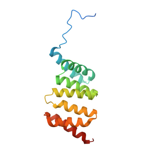
Type III secretion systems are a common virulence mechanism in many Gram-negative bacterial pathogens. These systems use a nanomachine resembling a molecular needle and syringe to provide an energized conduit for the translocation of effector proteins from the bacterial cytoplasm to the host cell cytoplasm for the benefit of the pathogen. Prior to translocation specialized chaperones maintain proper effector protein conformation. The class II chaperone, Invasion plasmid gene (Ipg) C, stabilizes two pore forming translocator proteins. IpgC exists as a functional dimer to facilitate the mutually exclusive binding of both translocators. In this study, we present the 3.3 A crystal structure of an amino-terminally truncated form (residues 10-155, denoted IpgC10-155) of the class II chaperone IpgC from Shigella flexneri. Our structure demonstrates an alternative quaternary arrangement to that previously described for a carboxy-terminally truncated variant of IpgC (IpgC1-151). Specifically, we observe a rotationally-symmetric "head-to- head" dimerization interface that is far more similar to that previously described for SycD from Yersinia enterocolitica than to IpgC1-151. The IpgC structure presented here displays major differences in the amino terminal region, where extended coil-like structures are seen, as opposed to the short, ordered alpha helices and asymmetric dimerization interface seen within IpgC1-151. Despite these differences, however, both modes of dimerization support chaperone activity, as judged by a copurification assay with a recombinant form of the translocator protein, IpaB. From primary to quaternary structure, these results presented here suggest that a symmetric dimerization interface is conserved across bacterial class II chaperones. In light of previous data which have described the structure and function of asymmetric dimerization, our results raise the possibility that class II chaperones may transition between asymmetric and symmetric dimers in response to changes in either biochemical modifications (e.g. proteolytic cleavage) or other biological cues. Such transitions may contribute to the broad range of protein-protein interactions and functions attributed to class II chaperones.
- Division of Cell Biology and Biophysics, School of Biological Sciences, University of Missouri-Kansas City, Kansas City, MO, USA.
Organizational Affiliation: