3KKX | pdb_00003kkx

Neutron structure of human carbonic anhydrase II


Experimental Data Snapshot

  • Method: NEUTRON DIFFRACTION
  • Resolution: 2.00 Å
  • R-Value Free: 
    0.286 (Depositor) 
  • R-Value Work: 
    0.275 (Depositor) 

Starting Model: experimental
View more details

wwPDB Validation 3D Report Full Report

Validation slider image for 3KKX

This is version 1.4 of the entry. See complete history

Literature

Neutron structure of human carbonic anhydrase II: implications for proton transfer.

Fisher, S.Z.Kovalevsky, A.Y.Domsic, J.F.Mustyakimov, M.McKenna, R.Silverman, D.N.Langan, P.A.

(2010) Biochemistry 49: 415-421

  • DOI: https://doi.org/10.1021/bi901995n
  • Primary Citation Related Structures: 
    3KKX

  • PubMed Abstract: 

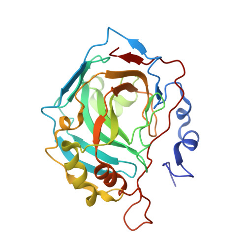
    Human carbonic anhydrase II (HCA II) catalyzes the reversible hydration of carbon dioxide to form bicarbonate and a proton. Despite many high-resolution X-ray crystal structures, mutagenesis, and kinetic data, the structural details of the active site, especially the proton transfer pathway, are unclear. A large HCA II crystal was prepared at pH 9.0 and subjected to vapor H-D exchange to replace labile hydrogens with deuteriums. Neutron diffraction studies were conducted at the Protein Crystallography Station at Los Alamos National Laboratory. The structure to 2.0 A resolution reveals several interesting active site features: (1) the Zn-bound solvent appearing to be predominantly a D(2)O molecule, (2) the orientation and hydrogen bonding pattern of solvent molecules in the active site cavity, (3) the side chain of His64 being unprotonated (neutral) and predominantly in an inward conformation pointing toward the zinc, and (4) the phenolic side chain of Tyr7 appearing to be unprotonated. The implications of these details are discussed, and a proposed mechanism for proton transfer is presented.


  • Organizational Affiliation
    • Bioscience Division MS M888, Los Alamos National Laboratory, Los Alamos, New Mexico 87544, USA. zfisher@lanl.gov

Macromolecule Content 

  • Total Structure Weight: 29.35 kDa 
  • Atom Count: 2,286 
  • Modeled Residue Count: 258 
  • Deposited Residue Count: 260 
  • Unique protein chains: 1

Macromolecules

Find similar proteins by:|  3D Structure
Entity ID: 1
MoleculeChains  Sequence LengthOrganismDetailsImage
Carbonic anhydrase 2260Homo sapiensMutation(s): 0 
Gene Names: CA2HCA II
EC: 4.2.1.1 (PDB Primary Data), 4.2.1.69 (UniProt)
UniProt & NIH Common Fund Data Resources
Find proteins for P00918 (Homo sapiens)
Explore P00918 
Go to UniProtKB:  P00918
PHAROS:  P00918
GTEx:  ENSG00000104267 
Entity Groups
Sequence Clusters30% Identity50% Identity70% Identity90% Identity95% Identity100% Identity
UniProt GroupP00918
Sequence Annotations
Expand
Reference Sequence

Small Molecules

Ligands 1 Unique
IDChains Name / Formula / InChI Key2D Diagram3D Interactions
ZN

Query on ZN


Download:Ideal Coordinates CCD File
B [auth A]ZINC ION
Zn
PTFCDOFLOPIGGS-UHFFFAOYSA-N

Experimental Data & Validation

Experimental Data

  • Method: NEUTRON DIFFRACTION
  • Resolution: 2.00 Å
  • R-Value Free:  0.286 (Depositor) 
  • R-Value Work:  0.275 (Depositor) 
Space Group: P 1 21 1
Unit Cell:
Length ( Å )Angle ( ˚ )
a = 42.6α = 90
b = 41.56β = 104.56
c = 72.82γ = 90
Software Package:
Software NamePurpose
PCSdata collection
nCNSrefinement
d*TREKdata reduction
SCALAdata scaling
nCNSphasing

Structure Validation

View Full Validation Report


Entry History 

Deposition Data

Revision History  (Full details and data files)

  • Version 1.0: 2010-01-12
    Type: Initial release
  • Version 1.1: 2011-07-13
    Changes: Version format compliance
  • Version 1.2: 2018-04-25
    Changes: Data collection
  • Version 1.3: 2018-06-20
    Changes: Data collection
  • Version 1.4: 2023-09-06
    Changes: Data collection, Database references, Derived calculations, Refinement description