Structural and Kinetic Evidence That Catalytic Reaction of Human UDP-glucose 6-Dehydrogenase Involves Covalent Thiohemiacetal and Thioester Enzyme Intermediates.
Egger, S., Chaikuad, A., Klimacek, M., Kavanagh, K.L., Oppermann, U., Nidetzky, B.(2012) J Biol Chem 287: 2119-2129
- PubMed: 22123821 Search on PubMedSearch on PubMed Central
- DOI: https://doi.org/10.1074/jbc.M111.313015
- Primary Citation Related Structures:
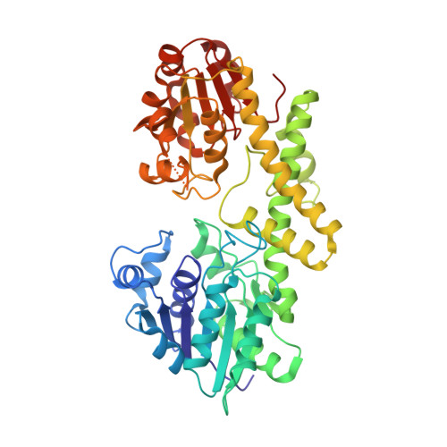
3KHU - PubMed Abstract:
Biosynthesis of UDP-glucuronic acid by UDP-glucose 6-dehydrogenase (UGDH) occurs through the four-electron oxidation of the UDP-glucose C6 primary alcohol in two NAD(+)-dependent steps. The catalytic reaction of UGDH is thought to involve a Cys nucleophile that promotes formation of a thiohemiacetal enzyme intermediate in the course of the first oxidation step. The thiohemiacetal undergoes further oxidation into a thioester, and hydrolysis of the thioester completes the catalytic cycle. Herein we present crystallographic and kinetic evidence for the human form of UGDH that clarifies participation of covalent catalysis in the enzymatic mechanism. Substitution of the putative catalytic base for water attack on the thioester (Glu(161)) by an incompetent analog (Gln(161)) gave a UGDH variant (E161Q) in which the hydrolysis step had become completely rate-limiting so that a thioester enzyme intermediate accumulated at steady state. By crystallizing E161Q in the presence of 5 mm UDP-glucose and 2 mm NAD(+), we succeeded in trapping a thiohemiacetal enzyme intermediate and determined its structure at 2.3 Å resolution. Cys(276) was covalently modified in the structure, establishing its role as catalytic nucleophile of the reaction. The thiohemiacetal reactive C6 was in a position suitable to become further oxidized by hydride transfer to NAD(+). The proposed catalytic mechanism of human UGDH involves Lys(220) as general base for UDP-glucose alcohol oxidation and for oxyanion stabilization during formation and breakdown of the thiohemiacetal and thioester enzyme intermediates. Water coordinated to Asp(280) deprotonates Cys(276) to function as an aldehyde trap and also provides oxyanion stabilization. Glu(161) is the Brønsted base catalytically promoting the thioester hydrolysis.
- Institute of Biotechnology and Biochemical Engineering, Graz University of Technology, A-8010 Graz, Austria.
Organizational Affiliation: