3KGK

Crystal structure of ArsD


Experimental Data Snapshot

  • Method: X-RAY DIFFRACTION
  • Resolution: 1.40 Å
  • R-Value Free: 0.219 
  • R-Value Work: 0.192 
  • R-Value Observed: 0.193 

wwPDB Validation   3D Report Full Report


This is version 1.3 of the entry. See complete history


Literature

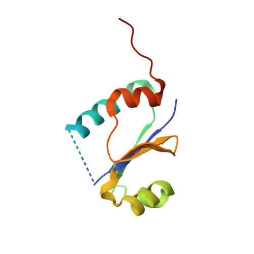
The 1.4 A crystal structure of the ArsD arsenic metallochaperone provides insights into its interaction with the ArsA ATPase.

Ye, J.Ajees, A.A.Yang, J.Rosen, B.P.

(2010) Biochemistry 49: 5206-5212

  • DOI: https://doi.org/10.1021/bi100571r
  • Primary Citation of Related Structures:  
    3KGK, 3MWH

  • PubMed Abstract: 

    Arsenic is a carcinogen that tops the Superfund list of hazardous chemicals. Bacterial resistance to arsenic is facilitated by ArsD, which delivers As(III) to the ArsA ATPase, the catalytic subunit of the ArsAB pump. Here we report the structure of the arsenic metallochaperone ArsD at 1.4 A and a model for its binding of metalloid. There are two ArsD molecules in the asymmetric unit. The overall structure of the ArsD monomer has a thioredoxin fold, with a core of four beta-strands flanked by four alpha-helices. Based on data from structural homologues, ArsD was modeled with and without bound As(III). ArsD binds one arsenic per monomer coordinated with the three sulfur atoms of Cys12, Cys13, and Cys18. Using this structural model, an algorithm was used to dock ArsD and ArsA. The resulting docking model provides testable predictions of the contact points of the two proteins and forms the basis for future experiments.


  • Organizational Affiliation

    Department of Biochemistry and Molecular Biology, Wayne State University School of Medicine, Detroit, Michigan 48201, USA.


Macromolecules
Find similar proteins by:  (by identity cutoff)  |  3D Structure
Entity ID: 1
MoleculeChains Sequence LengthOrganismDetailsImage
Arsenical resistance operon trans-acting repressor arsD
A, B
110Escherichia coliMutation(s): 3 
Gene Names: arsD
UniProt
Find proteins for P46003 (Escherichia coli)
Explore P46003 
Go to UniProtKB:  P46003
Entity Groups  
Sequence Clusters30% Identity50% Identity70% Identity90% Identity95% Identity100% Identity
UniProt GroupP46003
Sequence Annotations
Expand
  • Reference Sequence
Experimental Data & Validation

Experimental Data

  • Method: X-RAY DIFFRACTION
  • Resolution: 1.40 Å
  • R-Value Free: 0.219 
  • R-Value Work: 0.192 
  • R-Value Observed: 0.193 
  • Space Group: P 1 21 1
Unit Cell:
Length ( Å )Angle ( ˚ )
a = 37.841α = 90
b = 73.774β = 97.3
c = 41.248γ = 90
Software Package:
Software NamePurpose
ADSCdata collection
SOLVEphasing
REFMACrefinement
MOSFLMdata reduction
SCALAdata scaling

Structure Validation

View Full Validation Report


Entry History 

Deposition Data

Revision History  (Full details and data files)

  • Version 1.0: 2010-02-16
    Type: Initial release
  • Version 1.1: 2011-07-13
    Changes: Advisory, Version format compliance
  • Version 1.2: 2021-10-13
    Changes: Database references
  • Version 1.3: 2024-02-07
    Changes: Data collection, Database references