


Crystal Structure of Glucocerebrosidase Containing the N370S Mutation, Implications for Chaperon Therapy
Wei, R.R., Boucher, S., Hughes, H., Guziewica, N., Pan, C.Q., Edmunds, T.To be published.
Experimental Data Snapshot
Starting Model: experimental
View more details
Entity ID: 1 | |||||
|---|---|---|---|---|---|
| Molecule | Chains | Sequence Length | Organism | Details | Image |
| Glucosylceramidase | 497 | Homo sapiens | Mutation(s): 1 Gene Names: GBA, GC, GLUC EC: 3.2.1.45 (PDB Primary Data), 3.2.1.46 (UniProt), 2.4.1 (UniProt), 3.2.1 (UniProt) | 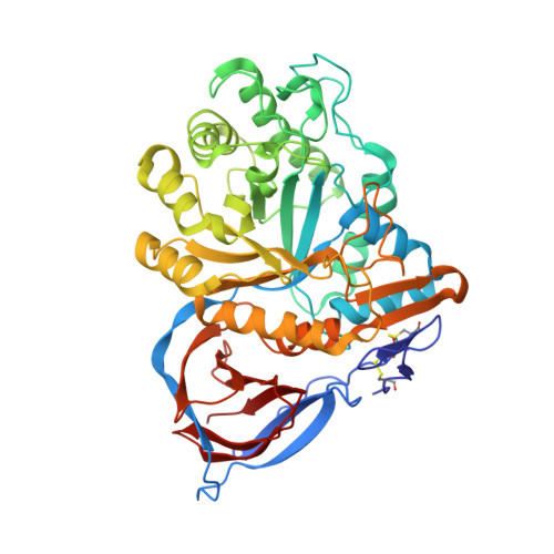 | |
UniProt & NIH Common Fund Data Resources | |||||
PHAROS: P04062 GTEx: ENSG00000177628 | |||||
Entity Groups | |||||
| Sequence Clusters | 30% Identity50% Identity70% Identity90% Identity95% Identity100% Identity | ||||
| UniProt Group | P04062 | ||||
Glycosylation | |||||
| Glycosylation Sites: 2 | Go to GlyGen: P04062-1 | ||||
Sequence AnnotationsExpand | |||||
Reference Sequence | |||||
Entity ID: 2 | |||||
|---|---|---|---|---|---|
| Molecule | Chains | Length | 2D Diagram | Glycosylation | D Interactions |
| 2-acetamido-2-deoxy-beta-D-glucopyranose-(1-4)-2-acetamido-2-deoxy-alpha-D-glucopyranose | C | 2 |  | N/A | |
Glycosylation Resources | |||||
GlyTouCan: G05026ZL GlyCosmos: G05026ZL GlyGen: G05026ZL | |||||
| Ligands 3 Unique | |||||
|---|---|---|---|---|---|
| ID | Chains | Name / Formula / InChI Key | 2D Diagram | 3D Interactions | |
| NAG Download:Ideal Coordinates CCD File | T [auth B] | 2-acetamido-2-deoxy-beta-D-glucopyranose C8 H15 N O6 OVRNDRQMDRJTHS-FMDGEEDCSA-N |  | ||
| SO4 Download:Ideal Coordinates CCD File | E [auth A] F [auth A] G [auth A] H [auth A] I [auth A] | SULFATE ION O4 S QAOWNCQODCNURD-UHFFFAOYSA-L |  | ||
| GOL Download:Ideal Coordinates CCD File | M [auth A], N [auth A], U [auth B] | GLYCEROL C3 H8 O3 PEDCQBHIVMGVHV-UHFFFAOYSA-N |  | ||
| Length ( Å ) | Angle ( ˚ ) |
|---|---|
| a = 109.465 | α = 90 |
| b = 285.064 | β = 90 |
| c = 92.225 | γ = 90 |
| Software Name | Purpose |
|---|---|
| HKL-2000 | data collection |
| MOLREP | phasing |
| REFMAC | refinement |
| MOSFLM | data reduction |
| SCALA | data scaling |


