3KCF | pdb_00003kcf

Crystal structure of TGFbRI complexed with a pyrazolone inhibitor


Experimental Data Snapshot

  • Method: X-RAY DIFFRACTION
  • Resolution: 2.80 Å
  • R-Value Free: 
    0.278 (Depositor), 0.273 (DCC) 
  • R-Value Work: 
    0.235 (Depositor), 0.232 (DCC) 
  • R-Value Observed: 
    0.237 (Depositor) 

Starting Model: experimental
View more details

wwPDB Validation 3D Report Full Report

Validation slider image for 3KCF

Ligand Structure Quality Assessment 


This is version 1.3 of the entry. See complete history

Literature

Pyrazolone based TGFbetaR1 kinase inhibitors.

Guckian, K.Carter, M.B.Lin, E.Y.Choi, M.Sun, L.Boriack-Sjodin, P.A.Chuaqui, C.Lane, B.Cheung, K.Ling, L.Lee, W.C.

(2010) Bioorg Med Chem Lett 20: 326-329

  • DOI: https://doi.org/10.1016/j.bmcl.2009.10.108
  • Primary Citation Related Structures: 
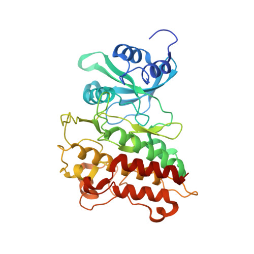
    3KCF

  • PubMed Abstract: 

    Interruption of TGFbeta signaling through inhibition of the TGFbetaR1 kinase domain may prove to have beneficial effect in both fibrotic and oncological diseases. Herein we describe the SAR of a novel series of TGFbetaR1 kinase inhibitors containing a pyrazolone core. Most TGFbetaR1 kinase inhibitors described to date contain a core five-membered ring bearing N as H-bond acceptor. Described herein is a novel strategy to replace the core structure with pyrazolone ring, in which the carbonyl group is designed as an H-bond acceptor to interact with catalytic Lys 232.


  • Organizational Affiliation
    • Biogen Idec Inc., 14 Cambridge Center, Cambridge, MA 02142, USA. kevin.guckian@biogenidec.com

Macromolecule Content 

  • Total Structure Weight: 197.64 kDa 
  • Atom Count: 13,297 
  • Modeled Residue Count: 1,642 
  • Deposited Residue Count: 1,710 
  • Unique protein chains: 1

Macromolecules

Find similar proteins by:|  3D Structure
Entity ID: 1
MoleculeChains  Sequence LengthOrganismDetailsImage
TGF-beta receptor type-1
A, B, C, D, E
342Homo sapiensMutation(s): 0 
Gene Names: TGFBR1TGFBRI
EC: 2.7.11.30
UniProt & NIH Common Fund Data Resources
Find proteins for P36897 (Homo sapiens)
Explore P36897 
Go to UniProtKB:  P36897
PHAROS:  P36897
GTEx:  ENSG00000106799 
Entity Groups
Sequence Clusters30% Identity50% Identity70% Identity90% Identity95% Identity100% Identity
UniProt GroupP36897
Sequence Annotations
Expand
Reference Sequence

Small Molecules

Ligands 2 Unique
IDChains Name / Formula / InChI Key2D Diagram3D Interactions
JZO

Query on JZO


Download:Ideal Coordinates CCD File
F [auth A],
J [auth B],
N [auth C],
P [auth D],
T [auth E]
4-[3-(methoxymethyl)phenyl]-1,2-dimethyl-5-quinoxalin-6-yl-1,2-dihydro-3H-pyrazol-3-one
C21 H20 N4 O2
OWJLLMXRMBEWIM-UHFFFAOYSA-N
PO4

Query on PO4


Download:Ideal Coordinates CCD File
G [auth A]
H [auth A]
I [auth A]
K [auth B]
L [auth B]
G [auth A],
H [auth A],
I [auth A],
K [auth B],
L [auth B],
M [auth B],
O [auth C],
Q [auth D],
R [auth D],
S [auth D],
U [auth E],
V [auth E],
W [auth E]
PHOSPHATE ION
O4 P
NBIIXXVUZAFLBC-UHFFFAOYSA-K
Binding Affinity Annotations 
IDSourceBinding Affinity
JZO BindingDB:  3KCF Ki: 35 (nM) from 1 assay(s)
IC50: 3690 (nM) from 1 assay(s)

Experimental Data & Validation

Experimental Data

  • Method: X-RAY DIFFRACTION
  • Resolution: 2.80 Å
  • R-Value Free:  0.278 (Depositor), 0.273 (DCC) 
  • R-Value Work:  0.235 (Depositor), 0.232 (DCC) 
  • R-Value Observed: 0.237 (Depositor) 
Space Group: C 2 2 21
Unit Cell:
Length ( Å )Angle ( ˚ )
a = 174.025α = 90
b = 249.076β = 90
c = 138.011γ = 90
Software Package:
Software NamePurpose
CBASSdata collection
CNXrefinement
HKL-2000data reduction
SCALEPACKdata scaling
CNXphasing

Structure Validation

View Full Validation Report


Ligand Structure Quality Assessment 


Entry History 

Deposition Data

Revision History  (Full details and data files)

  • Version 1.0: 2009-12-22
    Type: Initial release
  • Version 1.1: 2011-07-13
    Changes: Version format compliance
  • Version 1.2: 2024-02-21
    Changes: Data collection, Database references, Derived calculations
  • Version 1.3: 2024-04-03
    Changes: Refinement description