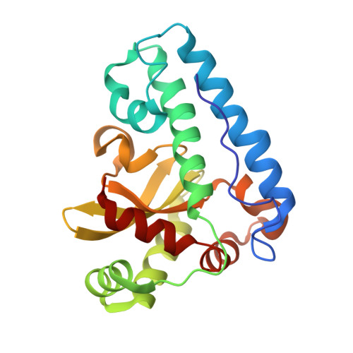
Structural Analysis of Peroxide-Soaked MnSOD Crystals Reveals Side-On Binding of Peroxide to Active-Site Manganese.
Porta, J., Vahedi-Faridi, A., Borgstahl, G.E.(2010) J Mol Biology 399: 377-384
- PubMed: 20417642 Search on PubMed
- DOI: https://doi.org/10.1016/j.jmb.2010.04.031
- Primary Citation Related Structures:
3K9S - PubMed Abstract:
The superoxide dismutase (SOD) enzymes are important antioxidant agents that protect cells from reactive oxygen species. The SOD family is responsible for catalyzing the disproportionation of superoxide radical to oxygen and hydrogen peroxide. Manganese- and iron-containing SOD exhibit product inhibition whereas Cu/ZnSOD does not. Here, we report the crystal structure of Escherichia coli MnSOD with hydrogen peroxide cryotrapped in the active site. Crystallographic refinement to 1.55 A and close inspection revealed electron density for hydrogen peroxide in three of the four active sites in the asymmetric unit. The hydrogen peroxide molecules are in the position opposite His26 that is normally assumed by water in the trigonal bipyramidal resting state of the enzyme. Hydrogen peroxide is present in active sites B, C, and D and is side-on coordinated to the active-site manganese. In chains B and D, the peroxide is oriented in the plane formed by manganese and ligands Asp167 and His26. In chain C, the peroxide is bound, making a 70 degrees angle to the plane. Comparison of the peroxide-bound active site with the hydroxide-bound octahedral form shows a shifting of residue Tyr34 towards the active site when peroxide is bound. Comparison with peroxide-soaked Cu/ZnSOD indicates end-on binding of peroxide when the SOD does not exhibit inhibition by peroxide and side-on binding of peroxide in the product-inhibited state of MnSOD.
- The Eppley Institute for Research in Cancer and Allied Diseases, 987696 Nebraska Medical Center, Omaha, NE 68198-7696, USA.
Organizational Affiliation: