A conserved motif within RAP1 has diversified roles in telomere protection and regulation in different organisms.
Chen, Y., Rai, R., Zhou, Z.R., Kanoh, J., Ribeyre, C., Yang, Y., Zheng, H., Damay, P., Wang, F., Tsujii, H., Hiraoka, Y., Shore, D., Hu, H.Y., Chang, S., Lei, M.(2011) Nat Struct Mol Biol 18: 213-221
- PubMed: 21217703 Search on PubMedSearch on PubMed Central
- DOI: https://doi.org/10.1038/nsmb.1974
- Primary Citation Related Structures:
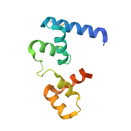
2L3N, 3K6G, 3OWT - PubMed Abstract:
Repressor activator protein 1 (RAP1) is the most highly conserved telomere protein. It is involved in protecting chromosome ends in fission yeast and promoting gene silencing in Saccharomyces cerevisiae, whereas it represses homology-directed recombination at telomeres in mammals. To understand how RAP1 has such diverse functions at telomeres, we solved the crystal or solution structures of the RAP1 C-terminal (RCT) domains of RAP1 from multiple organisms in complex with their respective protein-binding partners. Our analysis establishes RAP1(RCT) as an evolutionarily conserved protein-protein interaction module. In mammalian and fission yeast cells, this module interacts with TRF2 and Taz1, respectively, targeting RAP1 to chromosome ends for telomere protection. In contrast, S. cerevisiae RAP1 uses its RCT domain to recruit Sir3 to telomeres to mediate gene silencing. Together, our results show that, depending on the organism, the evolutionarily conserved RAP1 RCT motif has diverse functional roles at telomeres.
- Howard Hughes Medical Institute, University of Michigan Medical School, Ann Arbor, Michigan, USA.
Organizational Affiliation: