3JXB

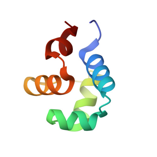
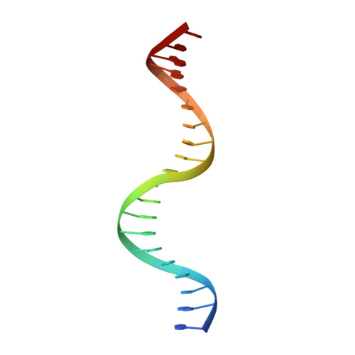
Crystal structure of the P22 c2 repressor protein in complex with synthetic operator 9C


Experimental Data Snapshot

  • Method: X-RAY DIFFRACTION
  • Resolution: 1.67 Å
  • R-Value Free: 0.226 
  • R-Value Work: 0.196 
  • R-Value Observed: 0.198 

Starting Model: experimental
View more details

wwPDB Validation   3D Report Full Report


This is version 1.2 of the entry. See complete history


Literature

Sequence Recognition of DNA by Protein-Induced Conformational Transitions

Watkins, D.Mohan, S.Koudelka, G.B.Williams, L.D.

(2010) J Mol Biol 396: 1145-1164

  • DOI: https://doi.org/10.1016/j.jmb.2009.12.050
  • Primary Citation of Related Structures:  
    3JXB, 3JXC, 3JXD

  • PubMed Abstract: 

    The binding of proteins to specific sequences of DNA is an important feature of virtually all DNA transactions. Proteins recognize specific DNA sequences using both direct readout (sensing types and positions of DNA functional groups) and indirect readout (sensing DNA conformation and deformability). Previously we showed that the P22 c2 repressor N-terminal domain (P22R NTD) forces the central non-contacted 5'-ATAT-3' sequence of the DNA operator into the B' state, a state known to affect DNA hydration, rigidity and bending. Usually the B' state, with a narrow minor groove and a spine of hydration, is reserved for A-tract DNA (TpA steps disrupt A-tracts). Here, we have co-crystallized P22R NTD with an operator containing a central 5'-ACGT-3' sequence in the non-contacted region. C.G base pairs have not previously been observed in the B' state and are thought to prevent it. However, P22R NTD induces a narrow minor groove and a spine of hydration to 5'-ACGT-3'. We observe that C.G base pairs have distinctive destabilizing and disordering effects on the spine of hydration. It appears that the reduced stability of the spine results in a higher energy cost for the B to B' transition. The differential effect of DNA sequence on the barrier to this transition allows the protein to sense the non-contacted DNA sequence.


  • Organizational Affiliation

    School of Chemistry and Biochemistry, Georgia Institute of Technology, Atlanta, GA 30332-0400, USA.


Macromolecules

Find similar proteins by:  (by identity cutoff)  |  3D Structure
Entity ID: 3
MoleculeChains Sequence LengthOrganismDetailsImage
Repressor protein C2
C, D
67Lederbergvirus P22Mutation(s): 0 
Gene Names: C2
UniProt
Find proteins for P69202 (Salmonella phage P22)
Explore P69202 
Go to UniProtKB:  P69202
Entity Groups  
Sequence Clusters30% Identity50% Identity70% Identity90% Identity95% Identity100% Identity
UniProt GroupP69202
Sequence Annotations
Expand
  • Reference Sequence

Find similar nucleic acids by:  Sequence   |   3D Structure  

Entity ID: 1
MoleculeChains LengthOrganismImage
5'-D(*CP*AP*TP*TP*TP*AP*AP*GP*AP*CP*GP*TP*CP*TP*TP*AP*AP*AP*TP*A)-3'20N/A
Sequence Annotations
Expand
  • Reference Sequence

Find similar nucleic acids by:  Sequence   |   3D Structure  

Entity ID: 2
MoleculeChains LengthOrganismImage
5'-D(*TP*AP*TP*TP*TP*AP*AP*GP*AP*CP*GP*TP*CP*TP*TP*AP*AP*AP*TP*G)-3'20N/A
Sequence Annotations
Expand
  • Reference Sequence
Experimental Data & Validation

Experimental Data

  • Method: X-RAY DIFFRACTION
  • Resolution: 1.67 Å
  • R-Value Free: 0.226 
  • R-Value Work: 0.196 
  • R-Value Observed: 0.198 
  • Space Group: P 21 21 21
Unit Cell:
Length ( Å )Angle ( ˚ )
a = 40.602α = 90
b = 54.319β = 90
c = 114.112γ = 90
Software Package:
Software NamePurpose
REFMACrefinement
PDB_EXTRACTdata extraction
HKL-2000data collection
DENZOdata reduction
SCALEPACKdata scaling
MOLREPphasing

Structure Validation

View Full Validation Report


Entry History 

Deposition Data

Revision History  (Full details and data files)

  • Version 1.0: 2010-01-19
    Type: Initial release
  • Version 1.1: 2011-07-13
    Changes: Advisory, Version format compliance
  • Version 1.2: 2023-09-06
    Changes: Data collection, Database references, Refinement description