3JVV | pdb_00003jvv

Crystal Structure of P. aeruginosa PilT with bound AMP-PCP


Experimental Data Snapshot

  • Method: X-RAY DIFFRACTION
  • Resolution: 2.60 Å
  • R-Value Free: 
    0.291 (Depositor), 0.292 (DCC) 
  • R-Value Work: 
    0.244 (Depositor), 0.247 (DCC) 
  • R-Value Observed: 
    0.247 (Depositor) 

Starting Model: experimental
View more details

wwPDB Validation 3D Report Full Report

Validation slider image for 3JVV

Ligand Structure Quality Assessment 


This is version 1.2 of the entry. See complete history

Literature

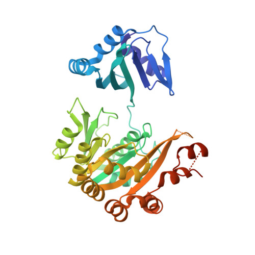
P. aeruginosa PilT structures with and without nucleotide reveal a dynamic type IV pilus retraction motor.

Misic, A.M.Satyshur, K.A.Forest, K.T.

(2010) J Mol Biology 400: 1011-1021

  • DOI: https://doi.org/10.1016/j.jmb.2010.05.066
  • Primary Citation Related Structures: 
    3JVU, 3JVV

  • PubMed Abstract: 

    Type IV pili are bacterial extracellular filaments that can be retracted to create force and motility. Retraction is accomplished by the motor protein PilT. Crystal structures of Pseudomonas aeruginosa PilT with and without bound beta,gamma-methyleneadenosine-5'-triphosphate have been solved at 2.6 A and 3.1 A resolution, respectively, revealing an interlocking hexamer formed by the action of a crystallographic 2-fold symmetry operator on three subunits in the asymmetric unit and held together by extensive ionic interactions. The roles of two invariant carboxylates, Asp Box motif Glu163 and Walker B motif Glu204, have been assigned to Mg(2+) binding and catalysis, respectively. The nucleotide ligands in each of the subunits in the asymmetric unit of the beta,gamma-methyleneadenosine-5'-triphosphate-bound PilT are not equally well ordered. Similarly, the three subunits in the asymmetric unit of both structures exhibit differing relative conformations of the two domains. The 12 degrees and 20 degrees domain rotations indicate motions that occur during the ATP-coupled mechanism of the disassembly of pili into membrane-localized pilin monomers. Integrating these observations, we propose a three-state "Ready, Active, Release" model for the action of PilT.


  • Organizational Affiliation
    • Department of Biomolecular Chemistry, University of Wisconsin-Madison, Madison, 1550 Linden Drive, Madison, WI 53706, USA.

Macromolecule Content 

  • Total Structure Weight: 120.02 kDa 
  • Atom Count: 7,939 
  • Modeled Residue Count: 996 
  • Deposited Residue Count: 1,068 
  • Unique protein chains: 1

Macromolecules

Find similar proteins by:|  3D Structure
Entity ID: 1
MoleculeChains  Sequence LengthOrganismDetailsImage
Twitching mobility protein
A, B, C
356Pseudomonas aeruginosaMutation(s): 0 
Gene Names: PA0395pilT
UniProt
Find proteins for P24559 (Pseudomonas aeruginosa (strain ATCC 15692 / DSM 22644 / CIP 104116 / JCM 14847 / LMG 12228 / 1C / PRS 101 / PAO1))
Explore P24559 
Go to UniProtKB:  P24559
Entity Groups
Sequence Clusters30% Identity50% Identity70% Identity90% Identity95% Identity100% Identity
UniProt GroupP24559
Sequence Annotations
Expand
Reference Sequence

Small Molecules

Ligands 3 Unique
IDChains Name / Formula / InChI Key2D Diagram3D Interactions
ACP

Query on ACP


Download:Ideal Coordinates CCD File
D [auth A],
G [auth B],
I [auth C]
PHOSPHOMETHYLPHOSPHONIC ACID ADENYLATE ESTER
C11 H18 N5 O12 P3
UFZTZBNSLXELAL-IOSLPCCCSA-N
CIT

Query on CIT


Download:Ideal Coordinates CCD File
F [auth A]CITRIC ACID
C6 H8 O7
KRKNYBCHXYNGOX-UHFFFAOYSA-N
MG

Query on MG


Download:Ideal Coordinates CCD File
E [auth A],
H [auth B],
J [auth C]
MAGNESIUM ION
Mg
JLVVSXFLKOJNIY-UHFFFAOYSA-N

Experimental Data & Validation

Experimental Data

  • Method: X-RAY DIFFRACTION
  • Resolution: 2.60 Å
  • R-Value Free:  0.291 (Depositor), 0.292 (DCC) 
  • R-Value Work:  0.244 (Depositor), 0.247 (DCC) 
  • R-Value Observed: 0.247 (Depositor) 
Space Group: C 2 2 21
Unit Cell:
Length ( Å )Angle ( ˚ )
a = 108.483α = 90
b = 119.552β = 90
c = 185.535γ = 90
Software Package:
Software NamePurpose
HKL-2000data collection
PHASERphasing
REFMACrefinement
HKL-2000data reduction
HKL-2000data scaling

Structure Validation

View Full Validation Report


Ligand Structure Quality Assessment 


Entry History 

Deposition Data

Revision History  (Full details and data files)

  • Version 1.0: 2010-06-02
    Type: Initial release
  • Version 1.1: 2011-07-13
    Changes: Advisory, Version format compliance
  • Version 1.2: 2023-09-06
    Changes: Data collection, Database references, Derived calculations, Refinement description